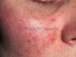

Rosacea affects mostly the central facial areas, including the nose, cheeks, brows, chin, and eyelids. It has classically been broken down into 4 descriptive subtypes: erythematotelangiectatic, papulopustular, phymatous, and ocular. As an individual may have combinations of and transitions between these subtypes, a new set of diagnostic criteria based on a more individualized phenotypic approach was developed and ratified by the Global ROSacea COnsensus (ROSCO) in 2017 and has been adopted by the American National Rosacea Society.
Either one of the following 2 findings are considered pathognomonic of rosacea:
- Phymatous change, which is due to fibrosis and/or sebaceous gland hyperplasia. This usually affects the nose (rhinophyma) but may occasionally manifest on the ears, chin, forehead, or eyelids.
- Persistent erythema of the central face that tends to flare in response to certain triggers.
- Flushing and/or temporary erythema. Common triggers include sun exposure, exposure to hot environments, stress, wind, physical exertion, alcohol consumption, and spicy foods.
- Papules and/or pustules.
- Telangiectasia, not limited to the alar areas.
- Burning, stinging, and dry sensations along with edema.
The etiology and pathogenic factors contributing to rosacea are still being delineated. The current consensus is that it results from an interplay between genetic predisposition, environmental factors, immune dysregulation, and neurovascular dysregulation.
Up to 40% of people with rosacea have an affected relative, and 2 genes have been found to be associated (HLA-DRA and BTNL2). Heat and stress have been found to stimulate the transient receptor channel vanilloid 1 (TRPV1), which has receptors on sensory neurons and endothelial cells. This leads to vasodilation and a burning sensation. Ultraviolet (UV) radiation directly upregulates matrix metalloproteins (MMPs), particularly MMP9, which influences collagenolysis, inflammation, and angiogenesis in the dermis. An increase in toll-like receptor 2 (TLR2) has been noted and is upregulated in the presence of Demodex overgrowth and the associated gram-negative Bacillus oleronius. TLR2 activates the cathelicidins pathway, producing both proinflammatory and angiogenic substances.
Additionally, topical steroids can cause rosacea-like eruptions, typically composed of monomorphic inflammatory papules and pustules. Topical calcineurin inhibitors may also be implicated.
Many rosacea associations have been reported in the literature, including cardiovascular (dyslipidemia, hypertension, coronary artery disease), gastrointestinal (Helicobacter pylori infection, ulcerative colitis), neurological (dementia, anxiety disorder, migraine), and immune disorders (rheumatoid arthritis, type 1 diabetes). Further investigation is warranted.

 Patient Information for