Contents

SynopsisCodesLook ForDiagnostic PearlsDifferential Diagnosis & PitfallsBest TestsManagement PearlsTherapyDrug Reaction DataReferences

Information for Patients

View all Images (13)

Rosacea - External and Internal Eye
See also in: Overview,Cellulitis DDx
Other Resources UpToDate PubMed Dermatology Online Journal

Rosacea - External and Internal Eye

See also in: Overview,Cellulitis DDx
Contributors: David O'Connell MD, Kimberley R. Zakka MD, MSc, Lauren Strazzula MD, Susan Burgin MD, Harvey A. Brown MD

Synopsis

Rosacea is a common, chronic inflammatory dermatosis that predominantly affects individuals with lighter skin colors, such as individuals of Northern European and Celtic descent. Onset is usually between ages 30-50 years, although it can affect all ages. Worldwide prevalence is between 3%-5% for adults. Women may be slightly more affected than men.

Rosacea affects mostly the central facial areas, including the nose, cheeks, brows, chin, and eyelids. It has classically been broken down into 4 descriptive subtypes: erythematotelangiectatic, papulopustular, phymatous, and ocular. As an individual may have combinations of and transitions between these subtypes, a new set of diagnostic criteria based on a more individualized phenotypic approach was developed and ratified by the Global ROSacea COnsensus (ROSCO) in 2017 and has been adopted by the American National Rosacea Society.

Either one of the following 2 findings are considered pathognomonic of rosacea:
  • Phymatous change, which is due to fibrosis and/or sebaceous gland hyperplasia. This usually affects the nose (rhinophyma) but may occasionally manifest on the ears, chin, forehead, or eyelids.
  • Persistent erythema of the central face that tends to flare in response to certain triggers.
There are 3 major features, the presence of any 2 of which is diagnostic of rosacea. All tend to involve the central face.
  • Flushing and/or temporary erythema. Common triggers include sun exposure, exposure to hot environments, stress, wind, physical exertion, alcohol consumption, and spicy foods.
  • Papules and/or pustules.
  • Telangiectasia, not limited to the alar areas.
Minor features:
  • Burning, stinging, and dry sensations along with edema.
Over 50% of all individuals with rosacea have some eye involvement, and it is the primary form of disease in approximately 20% of cases. Findings include lid margin telangiectasia, blepharitis, keratitis, conjunctivitis, and anterior uveitis. While there was limited ophthalmological representation on the ROSCO panel, it was thought that minimal diagnostic requirements for ocular rosacea would be a combination of lid margin telangiectasia and interpapillary injection or corneal abnormalities and scleral inflammation.

There is a 4-point grading scale for the severity of ocular rosacea:
  1. Mild – mild blepharitis and lid margin telangiectasia
  2. Mild-to-moderate – blepharoconjunctivitis
  3. Moderate-to-severe – blepharokeratoconjunctivitis
  4. Severe – sclerokeratitis, anterior uveitis
Many rosacea associations have been reported in the literature, including cardiovascular (dyslipidemia, hypertension, coronary artery disease), gastrointestinal (Helicobacter pylori infection, ulcerative colitis), neurological (dementia, anxiety disorder, migraine), and immune disorders (rheumatoid arthritis, type 1 diabetes). Further investigation is warranted.

Codes

ICD10CM:
L71.9 – Rosacea, unspecified

SNOMEDCT:
398909004 – Rosacea

Look For

Subscription Required

Diagnostic Pearls

Subscription Required

Differential Diagnosis & Pitfalls

To perform a comparison, select diagnoses from the classic differential

Subscription Required

Best Tests

Subscription Required

Management Pearls

Subscription Required

Therapy

Subscription Required

Drug Reaction Data

Subscription Required

References

Subscription Required

Last Reviewed:07/09/2026
Last Updated:08/11/2026
Copyright © 2026 VisualDx®. All rights reserved.
Patient Information for Rosacea - External and Internal Eye
Print E-Mail Images (13)
Contributors: Medical staff writer
Premium Feature
VisualDx Patient Handouts
Available in the Elite package
  • Improve treatment compliance
  • Reduce after-hours questions
  • Increase patient engagement and satisfaction
  • Written in clear, easy-to-understand language. No confusing jargon.
  • Available in English and Spanish
  • Print out or email directly to your patient
Copyright © 2026 VisualDx®. All rights reserved.
Rosacea - External and Internal Eye
See also in: Overview,Cellulitis DDx
A medical illustration showing key findings of Rosacea (Erythematotelangitatic)
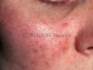
Clinical image of Rosacea - imageId=331974. Click to open in gallery.  caption: 'Erythema, telangiectasias, and few scattered inflammatory papules on the cheek.'
Erythema, telangiectasias, and few scattered inflammatory papules on the cheek.
Copyright © 2026 VisualDx®. All rights reserved.