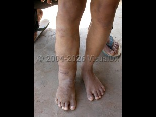

The global burden of lymphatic filariasis has declined substantially, with the estimated number of cases decreasing from 199 million in 2000 to approximately 50 million in 2018, largely due to widespread mass drug administration (MDA) programs. Between 2000 and 2023, the Global Programme to Eliminate Lymphatic Filariasis (GPELF) distributed more than 9.7 billion MDA doses to over 943 million people, reducing the at-risk population by 871 million. Nevertheless, the disease remains a significant public health concern. According to the World Health Organization (WHO), more than 657 million people across 39 countries remained at risk of lymphatic filariasis. The disease is endemic in many parts of Africa, Asia, South and Central America, and the Pacific Islands. More than two-thirds of infected people live in Asia, primarily in India. Risk factors include extended exposure to mosquitoes in endemic areas, especially near stagnant waters.
The prevalence of lymphatic filariasis increases with age, with most individuals in endemic areas exposed by adulthood. However, studies suggest that nearly one-third of children may acquire infection before age 5 years, often remaining asymptomatic except for possible lymphadenopathy until after puberty. The disease is more common in men than women.
Approximately 50% of infections are asymptomatic. The remainder of infections represent either acute lymphatic inflammation or chronic lymphatic obstruction. Travelers to endemic regions rarely develop chronic disease but may experience acute manifestations.
Acute lymphatic filariasis may present as:
- Acute adenolymphangitis, which is characterized by the sudden onset of fever, painful lymphadenopathy, and lymphangitis resulting from immune responses to dying adult worms.
- Acute dermatolymphangioadenitis, which is a bacterial complication of damaged skin, presenting with painful inflammatory plaques and systemic symptoms such as fever, chills, myalgia, and headache; recurrent episodes contribute to progression of lymphedema.
- Filarial fever, which consists of self-limiting febrile episodes that may occur without lymphatic inflammation.
- Tropical pulmonary eosinophilia, which results from hypersensitivity to microfilariae trapped in the lungs and typically manifests with nocturnal cough, wheezing, and marked eosinophilia.
- Lymphedema, the most common chronic manifestation, which results from progressive lymphatic dysfunction and typically affects the lower limbs, although the upper limbs, breasts, and genitalia may also be involved; advanced disease is known as elephantiasis.
- Genital involvement, which commonly manifests as unilateral or bilateral hydrocele. Rare involvement of the ovary, mesosalpinx, and vulva has also been reported.
- Renal complications, which include chyluria due to lymph leakage into the urinary tract. Proteinuria and hematuria may also occur.