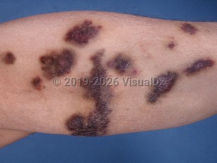
Kaposi sarcoma (KS) is a malignant neoplasm of lymphatic endothelial cell origin that occurs in several forms, including AIDS/HIV-associated KS, classic KS, African endemic KS, and iatrogenic KS. For discussion of classic, endemic, and iatrogenic forms of KS, see non-AIDS-associated Kaposi sarcoma. This summary discusses HIV-associated KS.
All types of KS are due to or influenced by human herpesvirus type 8 (HHV-8). Spindle cells of endothelial origin are the predominant cells affected. In the latent phase, HHV-8 antigens promote cell proliferation by inactivating the RB gene, which leads to transcription of S-phase genes and blocks apoptosis via p53 and p27Kip1 suppression. In the lytic phase, when tumor formation is noted, thousands of virion particles are assembled, resulting in cell lysis. HHV-8 requires additional cofactors for the development of KS. HIV coinfection acts as a stimulant for HHV-8 viral lytic expression and via its suppression of the immune system.
HIV-associated KS is the most common neoplasm in HIV-seropositive patients. This form of KS is primarily seen in the men who have sex with men (MSM) population, but it can be seen in female partners of men with the disease in addition to HIV-infected women. Lesions may worsen during immune reconstitution inflammatory syndrome and may also appear in patients with HIV who have received long-term antiretroviral therapy (ART). Patients with HIV-associated KS often have multifocal cutaneous disease. Additionally, HIV-associated KS is more likely than classic KS to display a rapidly progressive course.
The most common areas of involvement are the trunk and central face, especially the nose, and widespread involvement may be seen. In men, lesions on the penis are common. Oral involvement may occur in about one-third of KS cases and may be the initial manifestation of KS in about 15% of cases. Ophthalmic involvement, most often of the conjunctiva and eyelid, occurs in around one-fifth of patients with HIV-related KS.
Lesions in all forms of KS may progress to involve or be present in other organs such as the lymph nodes, lungs, gastrointestinal (GI) tract, liver, and spleen, but HIV-associated disease is more likely to have systemic involvement. This is more likely to occur in advanced stages of the disease and more aggressive disease variants. Most visceral cases are asymptomatic, but GI bleeding can occur.
The introduction of ART dramatically decreased the incidence, morbidity, and mortality of AIDS-associated KS.
AIDS-associated Kaposi sarcoma
See also in: External and Internal Eye,Anogenital,Oral Mucosal LesionAlerts and Notices
Important News & Links
Synopsis

Codes
ICD10CM:
C46.9 – Kaposi's sarcoma, unspecified
SNOMEDCT:
420524008 – Kaposi's sarcoma associated with AIDS
C46.9 – Kaposi's sarcoma, unspecified
SNOMEDCT:
420524008 – Kaposi's sarcoma associated with AIDS
Look For
Subscription Required
Diagnostic Pearls
Subscription Required
Differential Diagnosis & Pitfalls

To perform a comparison, select diagnoses from the classic differential
Subscription Required
Best Tests
Subscription Required
Management Pearls
Subscription Required
Therapy
Subscription Required
References
Subscription Required
Last Reviewed:08/10/2026
Last Updated:08/12/2026
Last Updated:08/12/2026
AIDS-associated Kaposi sarcoma
See also in: External and Internal Eye,Anogenital,Oral Mucosal Lesion