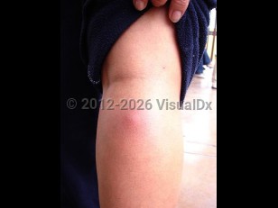
Cysticercosis refers to infection with the larval stage of the pork tapeworm Taenia solium. There is a frequent misconception that cysticercosis is acquired by eating pork; this is not the case. Human ingestion of pork containing larval cysts of T solium leads to taeniasis, a localized human intestinal adult tapeworm infection. Humans usually acquire cysticercosis via fecal-oral transmission of T solium eggs from tapeworm carriers via contaminated food or water. Once in the small intestine, the ingested eggs hatch and penetrate the mucosa. The larvae (oncospheres) then spread hematogenously throughout the body and encyst in various locations. The most frequently affected sites are skeletal muscle, subcutaneous tissue, the central nervous system (CNS), and ocular structures. Involvement of the heart, lungs, kidneys, liver, pancreas, and peritoneum have also been reported.
Cysticercosis is endemic to Mexico and Latin America, Eastern Europe, sub-Saharan Africa, Indonesia, China, and southeastern Asia. The World Health Organization (WHO) has included cysticercosis as one of the neglected tropical diseases. The incidence of this important parasitic disease is rising in the United States, especially in California and other states with a large immigrant population.
Clinical manifestations can begin months to years after acquisition. Subcutaneous cysticercosis manifests as multiple freely mobile nodules that rise years to decades after infestation. Its primary significance is as an indicator of possible neurocysticercosis, which will be present in around half of cases. Cysticerci may also invade skeletal muscle, giving rise to myositis (fever, myalgias, eosinophilia) and sometimes pseudohypertrophy. Intraocular cysticercosis may lead to vision loss.
Cysticercosis
Alerts and Notices
Important News & Links
Synopsis

Codes
ICD10CM:
B69.9 – Cysticercosis, unspecified
SNOMEDCT:
59051007 – Cysticercosis
B69.9 – Cysticercosis, unspecified
SNOMEDCT:
59051007 – Cysticercosis
Look For
Subscription Required
Diagnostic Pearls
Subscription Required
Differential Diagnosis & Pitfalls

To perform a comparison, select diagnoses from the classic differential
Subscription Required
Best Tests
Subscription Required
Management Pearls
Subscription Required
Therapy
Subscription Required
References
Subscription Required
Last Reviewed:07/30/2026
Last Updated:08/05/2026
Last Updated:08/05/2026
Cysticercosis