Oral melanotic macule
See also in: Oral Mucosal LesionAlerts and Notices
Important News & Links
Synopsis

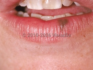
A benign hyperpigmentation of the mucous membranes. Presents as a solitary, flat, brown or grayish-brown discoloration of the lip, typically on the vermillion border, but may also occur on an intraoral mucosal surface. May be multiple. Appears slowly and has a uniform color and border. Typically 2-15 mm in diameter. In general, the darker the skin type, the more likely the patient is to develop mucosal melanotic macules. Although benign, biopsy may be needed to rule out a diagnosis of melanoma.
Codes
ICD10CM:
K13.79 – Other lesions of oral mucosa
SNOMEDCT:
235041006 – Oral melanocytic macule
K13.79 – Other lesions of oral mucosa
SNOMEDCT:
235041006 – Oral melanocytic macule
References
Subscription Required
Last Updated:02/09/2022
 Patient Information for Oral melanotic macule
Premium Feature
VisualDx Patient Handouts
Available in the Elite package
- Improve treatment compliance
- Reduce after-hours questions
- Increase patient engagement and satisfaction
- Written in clear, easy-to-understand language. No confusing jargon.
- Available in English and Spanish
- Print out or email directly to your patient
Upgrade Today

Oral melanotic macule
See also in: Oral Mucosal Lesion