Contents

SynopsisCodesLook ForDiagnostic PearlsDifferential Diagnosis & PitfallsBest TestsManagement PearlsTherapyDrug Reaction DataReferences

Information for Patients

View all Images (14)

Achilles tendonitis
Other Resources UpToDate PubMed

Achilles tendonitis

Contributors: Robert Lachky MD, Eric Ingerowski MD, FAAP
Other Resources UpToDate PubMed

Synopsis

Achilles tendonitis (or tendinitis) is an injury that describes inflammation and/or degeneration of the Achilles tendon. Patients typically present with posterior heel pain, swelling, burning, warmth, and/or stiffness.

Achilles tendinosis / tendinopathy is tendon thickening in the region approximately 2-6 cm proximal to tendon insertion and is thought to be caused by poor blood supply and anaerobic degeneration of the cartilage.

Insertional Achilles tendonitis is pain and tendon thickening at insertion of the Achilles tendon, wherein repetitive trauma leads to inflammation followed by cartilaginous, bony metaplasia.

Haglund deformity is enlargement of the posterosuperior tuberosity of calcaneus. Retrocalcaneal bursitis is inflammation of the space between the anterior aspect of Achilles tendon and posterior aspect of calcaneus.

Middle-aged or elderly patients are more likely to present with insertional Achilles tendonitis. Young, active patients are more likely to present with Haglund deformity or retrocalcaneal bursitis, which are often components of Achilles tendonitis.

Predisposing factors include overuse, imbalance of dorsiflexors / plantar flexors, use of fluoroquinolones, genetic predisposition, history of an inflammatory arthropathy (eg, gout), and poor tendon blood supply.

In addition to heel pain, history and physical examination findings may include:
  • Pain that worsens with activity, running
  • Shoe wear pain due to direct pressure
  • Pain with palpation (at insertion site of Achilles tendon if insertional tendonitis; there may be more fullness off to sides of tendon if retrocalcaneal bursitis and more proximal if Achilles tendinopathy)
  • Bony prominence at Achilles insertion
Related topic: Tendonitis

Codes

ICD10CM:
M76.60 – Achilles tendinitis, unspecified leg

SNOMEDCT:
11654001 – Achilles tendinitis

Look For

Subscription Required

Diagnostic Pearls

Subscription Required

Differential Diagnosis & Pitfalls

Best Tests

Subscription Required

Management Pearls

Subscription Required

Therapy

Subscription Required

Drug Reaction Data

Below is a list of drugs with literature evidence indicating an adverse association with this diagnosis. The list is continually updated through ongoing research and new medication approvals. Click on Citations to sort by number of citations or click on Medication to sort the medications alphabetically.

Subscription Required

References

Subscription Required

Last Reviewed:03/20/2018
Last Updated:06/25/2018
Copyright © 2023 VisualDx®. All rights reserved.
Patient Information for Achilles tendonitis
Print E-Mail Images (14)
Contributors: Medical staff writer

Overview

The Achilles tendon attaches the lower calf muscles to the heel bone. Achilles tendonitis, also called Achilles tendinitis, is a common overuse injury that can result from a sudden increase in running or exercise in people of all ages. The main cause of Achilles tendonitis is repetitive strain on the tendon. In most cases, this inflammation can be treated at home, however, when a tear or rupture occurs, more serious treatment is necessary.

Who’s At Risk

Increased risk of developing Achilles tendonitis:
  • Male sex (six times more likely than female)
  • Older adults who overdo exercise and sports without adequate preparation
  • Foot abnormalities (flat foot, high arch, leg-length discrepancy)
  • Use of worn running shoes
  • Repetitive motions, extensive cold-weather running, and on steep terrain
  • High blood pressure, or psoriasis
  • Certain prescription drugs, such as glucocorticoids

Signs & Symptoms

Achilles tendonitis typically begins with an ache in the back of the leg above the heel after exercise. If left untreated, pain begins to become more severe. Your Achilles tendon may also become tender or stiff in the morning.

Self-Care Guidelines

Follow the RICE guidelines to help treat symptoms:
  • Rest for several days to limit strain on the Achilles tendon
  • To decrease swelling and pain, ice for 15 minutes after exercise or when you are experiencing pain
  • You can compress the tendon by wearing elastic bandages around the tendon
  • Elevate the foot above your heart when sleeping or resting to reduce swelling

To prevent Achilles tendonitis, start exercise regimens gradually and slowly build up to your higher levels to reduce strain on the Achilles tendon. Make sure that you do not overuse running shoes because old shoes can put extra strain on the Achilles tendon. You can also help prevent Achilles tendonitis by strengthening your calf muscles with daily exercises such as calf raises.

If symptoms persist, cross train with low impact exercises a few days per week such as aqua jogging, swimming, or cycling instead of high impact exercises.

When to Seek Medical Care

If you experience pain or tenderness around the Achilles tendon, contact a health care provider.

Treatments

If over-the-counter medication such as ibuprofen (Advil) are not sufficient, you may be given stronger medications to help reduce inflammation.

A physical therapist may prescribe stretching exercises to help strengthen and heal the Achilles tendon. Also, you may be given orthotic devices in your shoes to elevate your heel. Orthotics help to cushion the heel and limit the amount of stress on your Achilles tendon.

If conservative treatments mentioned above do not improve the pain, your health care provider may recommend surgery.
Copyright © 2023 VisualDx®. All rights reserved.
Achilles tendonitis
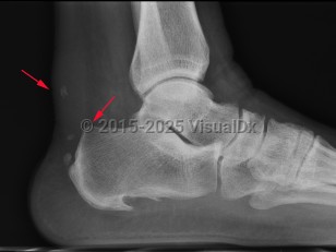
Imaging Studies image of Achilles tendonitis - imageId=7886591. Click to open in gallery.  caption: '<span>Achilles Tendonitis - rupture.  Lateral ankle radiograph demonstrates soft tissue thickening and  multiple, small dystrophic calcifications involving the distal Achilles  tendon compatible with chronic Achilles tendonitis. Note the presence of  a small bony bump along the posterosuperior calcaneus which may be the  early signs of a Haglund deformity.</span>'
Achilles Tendonitis - rupture. Lateral ankle radiograph demonstrates soft tissue thickening and multiple, small dystrophic calcifications involving the distal Achilles tendon compatible with chronic Achilles tendonitis. Note the presence of a small bony bump along the posterosuperior calcaneus which may be the early signs of a Haglund deformity.
Copyright © 2023 VisualDx®. All rights reserved.