Meningococcal disease is a rapidly progressive infection caused by Neisseria meningitides, a Gram-negative diplococcus bacterium. Infection begins as a nonspecific viral-like illness that rapidly evolves (within hours) into one of two main presentations: meningitis or septicemia. Apart from epidemics, meningococcal disease occurs sporadically, most commonly during the winter months, often following outbreaks of influenza. Most cases are acquired through exposure to asymptomatic carriers via respiratory droplets. Although severe disease has a mortality approaching 50%, early recognition and aggressive management can reduce the mortality to less than 5%.
Complications of acute meningococcemia include pericarditis / myocarditis, disseminated intravascular coagulation (DIC), meningitis and permanent neurologic sequelae, septic arthritis, osteomyelitis, adrenal hemorrhage (Waterhouse-Friderichsen syndrome), gangrene, and death.
Risk factors for meningococcal disease include viral infections, smoke exposure, crowded living conditions, underlying chronic diseases, and low socioeconomic status. Infants with primary or acquired deficiencies of terminal complement components or asplenia are also at increased risk of meningococcal disease.
Related topic: chronic meningococcemia
Potentially life-threatening emergency
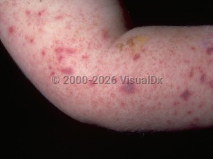
Acute meningococcemia in Infant/Neonate
Alerts and Notices
Important News & Links
Synopsis

Codes
ICD10CM:
A39.2 – Acute meningococcemia
SNOMEDCT:
186365005 – Acute meningococcemia
A39.2 – Acute meningococcemia
SNOMEDCT:
186365005 – Acute meningococcemia
Look For
Subscription Required
Diagnostic Pearls
Subscription Required
Differential Diagnosis & Pitfalls

To perform a comparison, select diagnoses from the classic differential
Subscription Required
Best Tests
Subscription Required
Management Pearls
Subscription Required
Therapy
Subscription Required
Drug Reaction Data
Subscription Required
References
Subscription Required
Last Updated:02/18/2024
Potentially life-threatening emergency
Acute meningococcemia in Infant/Neonate