Age-related macular degeneration (AMD) is a degenerative disorder affecting people 50 and older that may impair central vision. AMD is now the leading cause of permanent blindness in the Western world. The incidence increases with age. The disease is more common in white individuals, females, and those with a positive family history of AMD. Obesity, smoking, hyperopia, light iris color, hypertension, high cholesterol, and cardiovascular disease are also contributing factors.
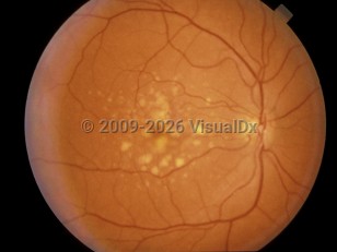
With age, the retinal pigment epithelium (RPE) accumulates waste products from the retina. Drusen, the hallmark of AMD, are focal deposits of this debris between the RPE and Bruch's membrane. Excess drusen can damage the RPE, leading to retinal atrophy and the expression of angiogenic cytokines. With choroidal neovascularization (CNV), choroidal vessels grow abnormally and leak or bleed into the subretinal space through defects in Bruch's membrane.
AMD can be divided into a non-neovascular (dry) atrophic type and an exudative neovascular (wet) type. The atrophic form comprises 90% of AMD and only 10% of blindness, while the exudative form makes up 10% of AMD but is responsible for 90% of vision loss.
Patients with early AMD are often asymptomatic. As the drusen cover more of the macula, patients may complain of fluctuating blurry vision or loss of contrast sensitivity. With exudative AMD, patients often note visual distortion or loss of vision.
Age-related macular degeneration - External and Internal Eye
Alerts and Notices
Important News & Links
Synopsis

Codes
ICD10CM:
H35.30 – Unspecified macular degeneration
SNOMEDCT:
267718000 – Age-Related Macular Degeneration
H35.30 – Unspecified macular degeneration
SNOMEDCT:
267718000 – Age-Related Macular Degeneration
Look For
Subscription Required
Diagnostic Pearls
Subscription Required
Differential Diagnosis & Pitfalls

To perform a comparison, select diagnoses from the classic differential
Subscription Required
Best Tests
Subscription Required
Management Pearls
Subscription Required
Therapy
Subscription Required
References
Subscription Required
Last Updated:07/23/2013
Age-related macular degeneration - External and Internal Eye