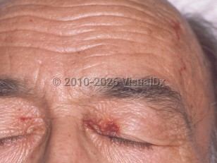
Amyloidosis is caused by extracellular deposition of polymerized protein fibrils in various tissues and organs. Amyloid is organized into beta-plated sheets and, when viewed stained with Congo red dye and then viewed with polarized light, has apple-green birefringence. Amyloidosis may be limited to the skin (eg, lichen amyloidosis) or be systemic. The systemic type may be primary (AL amyloidosis) or secondary (AA amyloidosis). Amyloid proteins may deposit in almost any organ. Cutaneous manifestations are seen in 50% of patients with AL amyloidosis and may present with petechiae, ecchymoses, and purpura, particularly in the flexural areas, periorbital area, and sites of trauma. Other cutaneous signs are nodules or plaques, waxy papules, hypo- or hyperpigmentation, skin thickening, scleroderma-like changes, and bullae.
Nail changes in amyloidosis are much less common than cutaneous manifestations. Nail dystrophy is secondary to nail matrix and bed amyloid deposition leading to faulty nail plate production. Nail dystrophy usually slowly worsens over time. In about one-half of cases, cutaneous manifestations precede nail changes. In about 25% of cases, nail changes may be the presenting sign of amyloidosis.
The most common underlying diagnosis in patients with nail changes is myeloma-associated systemic amyloidosis, followed by an underlying monoclonal gammopathy; in some cases, amyloidosis is idiopathic.
For nail dystrophy due to amyloidosis, there is a slight male predominance. It usually presents in the sixth or seventh decade (range: 49-81 years).
The severity of cardiac dysfunction at the time of diagnosis impacts the survival of patients with systemic AL amyloidosis. Patients without cardiac impairment survive for many years, while patients who are diagnosed after advanced heart damage have a median survival of 3-6 months.
Related topics: multiple myeloma, monoclonal gammopathy
AL amyloidosis - Nail and Distal Digit
See also in: Overview,External and Internal Eye,Oral Mucosal LesionAlerts and Notices
Important News & Links
Synopsis

Codes
ICD10CM:
E85.81 – Light chain (AL) amyloidosis
SNOMEDCT:
23132008 – AL amyloidosis
E85.81 – Light chain (AL) amyloidosis
SNOMEDCT:
23132008 – AL amyloidosis
Look For
Subscription Required
Diagnostic Pearls
Subscription Required
Differential Diagnosis & Pitfalls

To perform a comparison, select diagnoses from the classic differential
Subscription Required
Best Tests
Subscription Required
Management Pearls
Subscription Required
Therapy
Subscription Required
References
Subscription Required
Last Reviewed:10/14/2019
Last Updated:10/02/2024
Last Updated:10/02/2024
AL amyloidosis - Nail and Distal Digit
See also in: Overview,External and Internal Eye,Oral Mucosal Lesion