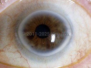
Arcus senilis - External and Internal Eye
Alerts and Notices
Important News & Links
Synopsis

Arcus senilis is associated with lipid deposition within the corneal stroma. It manifests clinically as annular blue, grey, or white deposits within the periphery of the cornea with a distinct clear perilimbic zone. Its prevalence increases with age. Early onset may be associated with hypercholesterolemia. The same condition in younger patients is known as arcus juvenilis.
Codes
ICD10CM:
H18.419 – Arcus senilis, unspecified eye
SNOMEDCT:
18735004 – Arcus senilis
H18.419 – Arcus senilis, unspecified eye
SNOMEDCT:
18735004 – Arcus senilis
Look For
Subscription Required
Diagnostic Pearls
Subscription Required
Differential Diagnosis & Pitfalls

To perform a comparison, select diagnoses from the classic differential
Subscription Required
Best Tests
Subscription Required
Management Pearls
Subscription Required
Therapy
Subscription Required
References
Subscription Required
Last Updated:01/11/2022
Arcus senilis - External and Internal Eye

