Contents

SynopsisCodesLook ForDiagnostic PearlsDifferential Diagnosis & PitfallsBest TestsManagement PearlsTherapyReferences

Information for Patients

View all Images (87)

Atopic dermatitis in Infant/Neonate
See also in: Cellulitis DDx,External and Internal Eye
Other Resources UpToDate PubMed

Atopic dermatitis in Infant/Neonate

See also in: Cellulitis DDx,External and Internal Eye
Contributors: Azeen Sadeghian MD, Sophia Delano MD, Susan Burgin MD
Other Resources UpToDate PubMed

Synopsis

Atopic dermatitis (eczema) is a chronic, relapsing, pruritic condition characterized by (1) pruritus (itch); (2) facial and extensor involvement during infancy that changes to flexural involvement in late childhood; (3) a chronic relapsing course; and (4) a personal or family history of atopy (atopic dermatitis, food allergies, allergic rhinitis, and/or asthma).

Infants and children are most often affected, with 85% of cases appearing in the first year of life, and 95% of cases appearing by 5 years. Uncommonly, the condition may persist into, or even arise in, adulthood. Less than 1% of adults are affected by atopic dermatitis.

In infants, the disease involves primarily the face, scalp, torso, and extensor aspects of extremities. In children and adults, the disease usually involves chiefly the flexural aspects of extremities, but it may be more generalized. In adults, flexural skin may be clear, and disease may be focal or widespread. Follicular patterns of atopic dermatitis (ie, follicular eczema) are more common in persons with darker skin phototypes.

Atopic dermatitis may be categorized as follows:
  • Acute – erythema, vesicles, bullae, weeping, crusting
  • Subacute – scaly plaques, papules, round erosions, crusts
  • Chronic eczema – lichenification, scaling, hyper- and hypopigmentation
The cause of atopic dermatitis is unknown. Genetic and environmental predisposing factors exist. Multiple loci have been associated with atopic dermatitis. A family history of atopic dermatitis is common.

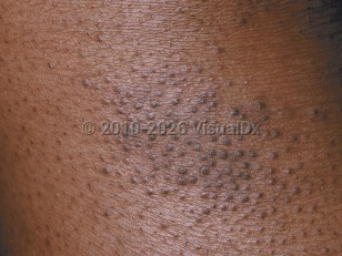
Intense pruritus is a hallmark of atopic dermatitis. Scratching leads to lichenification (skin thickening). Impaired barrier function results in increased transepidermal water loss and susceptibility to secondary bacterial and viral infections. Patients with atopic dermatitis are prone to impetiginization with Staphylococcus aureus. Secondary infections with herpes simplex virus (eczema herpeticum), Coxsackie viruses (eczema coxsackium), vaccinia virus (eczema vaccinatum), or molluscum contagiosum may occur.

Patients with atopic dermatitis have difficulties in retaining skin moisture and suffer from xerosis (dry skin). Environmental triggers, such as heat, humidity, detergents / soaps, abrasive clothing, chemicals, smoke, and even stress, tend to aggravate the condition. Latex allergy and nickel allergy occur more often in persons with atopic dermatitis. Additionally, patients with atopic dermatitis have been found to be more likely to have positive patch test results to products commonly found in topical treatments, including cocamidopropyl betaine, wool alcohol / lanolin, and tixocortol pivalate. Allergy to eggs, cow's milk, or peanuts is common. There may be a relationship between atopic dermatitis and the development of aspirin-related respiratory disease.

Atopic dermatitis is increasing in the developed world. In the United States, about 10% of children may be affected by atopic dermatitis, but the majority of these cases are mild.

Codes

ICD10CM:
L20.9 – Atopic dermatitis, unspecified

SNOMEDCT:
24079001 – Atopic dermatitis

Look For

Subscription Required

Diagnostic Pearls

Subscription Required

Differential Diagnosis & Pitfalls

  • Infantile seborrheic dermatitis tends to involve the scalp and groin and has greasy scale as opposed to dry scale seen in atopic dermatitis. Infants with atopic dermatitis often have seborrheic dermatitis as well, and there can be overlap between the conditions.
  • The impaired cutaneous barrier in patients with atopic dermatitis makes them more prone to irritant contact dermatitis. Allergic contact dermatitis is also frequently encountered in atopic patients.
  • Langerhans cell histiocytosis presents as petechial, eroded papules of the scalp, axilla, hands, feet, or groin with associated lymphadenopathy.
  • Scabies is also intensely pruritic, classically accentuated at night. Typical sites of involvement include the interdigital web spaces, axillae, wrists, belt area, buttocks, and feet. The pathognomic sign is the burrow. In young children, scabies may present as widespread, pruritic, crusted vesicopustules.
  • Lesions in tinea corporis are typically annular or arcuate. Tinea incognito may be mistaken for atopic dermatitis due to absence of scale and inflammation. Fungal elements can be demonstrated using a potassium hydroxide (KOH) preparation.
  • Classic lesions in psoriasis are well-defined erythematous plaques involving the scalp and extensor elbows and knees with overlying silvery scale. In infants and toddlers, psoriasis often presents as well-dermarcated, pink plaques in the groin that involve the intertriginous folds. Groin psoriasis in young children typically improves once the child is potty-trained.
  • An atopic-like eczematous dermatitis may be observed in patients with genetic immunodeficiencies (eg, Wiskott-Aldrich syndrome, hyperimmunoglobulinemia E syndrome, selective IgA deficiency, Omenn syndrome), often associated with failure to thrive, recurrent infections, hematologic abnormalities, and chronic diarrhea.
  • Disorders of keratinization (eg, nonbullous congenital ichthyosiform erythroderma, Netherton syndrome) may appear similar but present at birth rather than 3-6 months of age.
  • Nutritional deficiencies (eg, phenylketonuria, multiple carboxylase deficiency, zinc deficiency, essential fatty acid deficiency) may appear identical to atopic dermatitis but are usually associated with failure to thrive and systemic symptoms.

Best Tests

Subscription Required

Management Pearls

Subscription Required

Therapy

Subscription Required

References

Subscription Required

Last Reviewed:03/22/2017
Last Updated:11/01/2022
Copyright © 2023 VisualDx®. All rights reserved.
Patient Information for Atopic dermatitis in Infant/Neonate
Print E-Mail Images (87)
Contributors: Medical staff writer

Overview

Eczema (atopic dermatitis) is a common disorder in infants causing dry and itchy patches of skin. Infants may scratch at the dry skin, causing bleeding in the affected areas and sleep disruption. Eczema can occur on any part of the body. In infants, it is seen on the cheeks and on skin that is stretched often (eg, the back of elbows and front of knees).

Eczema is not contagious and it is not an infection, but prolonged scratching can allow bacteria to get through the skin and cause an infection. Some common triggers for eczema include:
  • Heat and humidity.
  • Abrasive clothing.
  • Tobacco smoke and chemicals.
  • Some soaps and detergents.

Who’s At Risk

Infants are more likely to have eczema if they have other allergies, such as to foods or milk, as well as seasonal allergies, or if they have other family members who were affected by eczema as an infant. Infants who have asthma are also more likely to have eczema.

Signs & Symptoms

The first location affected by eczema in an infant is usually the cheeks. The scalp, elbows and knees, and skin of other parts of the body may be affected as well.
  • Thickened, scaly papules (small, raised bumps) and plaques (areas of raised skin that are larger than a thumbnail and feel rough and flaky) are seen in these areas. The condition may be:
    • Mild – few, scattered areas of involvement, which are easily treated with self-care measures.
    • Moderate – more extensive involvement, which is more difficult to control with self-care measures and may require prescription therapy.
    • Severe – diffuse involvement, which is difficult to treat even with prescription therapy.
  • In lighter skin colors, affected areas may appear pink or red; in darker skin colors, the redness may be subtle, or affected areas may appear purplish or darker brown.
  • Areas of eczema that become infected can develop thick crusts.

Self-Care Guidelines

Maintaining healthy skin is very important for those with eczema.
  • Moisturizing skin-care routines are essential.
  • Hypoallergenic moisturizing soaps, such as unscented Dove Sensitive Skin Beauty Bar, Vanicream Cleansing Bar, and Nature by Canus, are recommended.
  • Thick moisturizers such as petroleum jelly (Vaseline), Aquaphor Healing Ointment, Eucerin Original Healing Cream, CeraVe Healing Ointment, or CeraVe Moisturizing Cream should be applied to damp skin daily after bathing.
  • Attempt to minimize exposure to heat, humidity, detergents / soaps, abrasive clothing, chemicals, smoke, and stress.
  • Fragrance-free laundry detergent may be beneficial.
  • Keep the home from getting too dry by using a humidifier, especially in the bedroom.

When to Seek Medical Care

See your child's medical professional for evaluation if you see no improvement with self-care measures or the eczema gets worse. Also seek medical care if you see areas of pus or large numbers of crusty areas (scabs), as this might be caused by infection with bacteria.

Treatments

Topical or oral medications for eczema can include:
  • Topical steroid creams or ointments to treat active areas of localized eczema. Lower-strength steroids may be used on the face, and medium-to-high-strength steroids may be used on the body (trunk) and arms or legs (extremities). Care should be taken when using topical steroids in skin folds and on covered (obstructed) skin areas due to the risk of thinning (atrophy) of the skin.
  • Oral antihistamines may be prescribed to decrease itching.
  • If an infection is suspected, topical or oral antibiotics may be prescribed.
  • In patients who have multiple areas of broken skin or a history of bacterial skin infections, dilute bleach baths may be prescribed.
Copyright © 2023 VisualDx®. All rights reserved.
Atopic dermatitis in Infant/Neonate
See also in: Cellulitis DDx,External and Internal Eye
A medical illustration showing key findings of Atopic dermatitis : Bilateral distribution, Dry skin, Erythema, Excoriated skin lesion, Flexural distribution, Lichenified plaque, Thickened skin, Ocular pruritus, Pruritus
Clinical image of Atopic dermatitis - imageId=214627. Click to open in gallery.  caption: 'A close-up of follicular papules with scale.'
A close-up of follicular papules with scale.
Copyright © 2023 VisualDx®. All rights reserved.