An atypical nevus is a benign melanocytic lesion that appears clinically atypical with asymmetry, large size, irregular or ill-defined borders, and/or varied coloration. A dysplastic nevus (also referred to as a Clark nevus) is a histopathologic term that describes a melanocytic proliferation showing architectural disorder and cytologic atypia and sharing some features of melanoma. Dysplastic nevi usually appear clinically atypical and atypical nevi may be dysplastic; however, the clinical and histopathologic terms do not always completely correlate.
There is controversy around the term dysplastic nevus, including a lack of consensus on how it is defined and what it represents biologically. Originally described in melanoma-prone families, dysplastic nevi were part of a clinical phenotype. Patients characteristically had numerous nevi, which often appeared clinically atypical; on biopsy (as initially described by Dr. Wallace Clark), 4 main histologic features were observed: 1) atypical melanocytic hyperplasia, 2) melanocytes with cytologic features characteristic of malignant melanocytes, 3) mesenchymal changes in the papillary dermis, and 4) lymphocytic infiltrate. Since then, dysplastic nevi have been recognized to occur spontaneously, and different variations of the above diagnostic criteria (including grading of cytologic atypia as mild, moderate, or severe) have been proposed without a clear diagnostic consensus.
Dysplastic nevi usually begin to appear in childhood through early adult years and have an estimated prevalence ranging from 2%-10%. They are thought to have a genetic component and are more common in phototype I-III individuals. While less prevalent than common nevi, dysplastic nevi are believed to correlate with the overall number of melanocytic lesions in an individual and are thought to confer a 4- to 15-fold increased risk of melanoma.
While there is an increased relative risk of melanoma in individuals with multiple dysplastic nevi and dysplastic nevi can share clinical and histologic features with melanoma, current evidence does not support a dysplastic nevus as a definite premalignant lesion. Studies have shown that most melanomas appear to arise de novo without an associated nevus, and even when an associated nevus is present in histologic contiguity with melanoma, it is more frequently a common nevus than a dysplastic nevus.
Related topic: agminated nevus
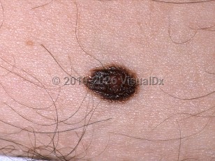
Atypical nevus - Hair and Scalp
See also in: OverviewAlerts and Notices
Important News & Links
Synopsis

Codes
ICD10CM:
D23.9 – Other benign neoplasm of skin, unspecified
SNOMEDCT:
254818000 – Atypical nevus of skin
D23.9 – Other benign neoplasm of skin, unspecified
SNOMEDCT:
254818000 – Atypical nevus of skin
Look For
Subscription Required
Diagnostic Pearls
Subscription Required
Differential Diagnosis & Pitfalls

To perform a comparison, select diagnoses from the classic differential
Subscription Required
Best Tests
Subscription Required
Management Pearls
Subscription Required
Therapy
Subscription Required
References
Subscription Required
Last Updated:02/09/2023
 Patient Information for Atypical nevus - Hair and Scalp
Premium Feature
VisualDx Patient Handouts
Available in the Elite package
- Improve treatment compliance
- Reduce after-hours questions
- Increase patient engagement and satisfaction
- Written in clear, easy-to-understand language. No confusing jargon.
- Available in English and Spanish
- Print out or email directly to your patient
Upgrade Today

Atypical nevus - Hair and Scalp
See also in: Overview