Contents

SynopsisCodesLook ForDiagnostic PearlsDifferential Diagnosis & PitfallsBest TestsManagement PearlsTherapyReferences

View all Images (8)

Benign salivary gland tumor - Oral Mucosal Lesion
Other Resources UpToDate PubMed

Benign salivary gland tumor - Oral Mucosal Lesion

Contributors: Carl Allen DDS, MSD, Sook-Bin Woo MS, DMD, MMSc
Other Resources UpToDate PubMed

Synopsis

Benign intraoral salivary gland tumors have no identifiable etiologic factors and consist mainly of the neoplasms known as pleomorphic adenoma and monomorphic adenoma. These tumors affect mainly adults, with a slight female predilection.

The pleomorphic adenoma intraorally involves the posterior hard palate or anterior soft palate most commonly, but can be seen in the buccal mucosa and labial mucosa. The upper labial mucosa is the most common site for the monomorphic adenoma.

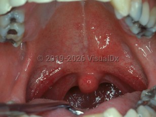
These neoplasms present as a unilateral slowly growing asymptomatic nodule. On palpation these neoplasms usually have a rubbery consistency and tumors involving the labial, buccal or soft palate mucosa are freely movable. Tumors of the posterior hard palate are not movable because they are set in the densely collagenous tissue of the hard palate.

A monomorphic adenoma with a prominent cystic component may appear bluish in color, but most lesions show no significant color change.

Such tumors may also develop in the major salivary glands.

Codes

ICD10CM:
D11.9 – Benign neoplasm of major salivary gland, unspecified

SNOMEDCT:
255154009 – Benign tumor of salivary gland

Look For

Subscription Required

Diagnostic Pearls

Subscription Required

Differential Diagnosis & Pitfalls

  • Dental abscess (see abscess, cellulitis) – Palatal extension of periapical inflammatory disease is seen occasionally and may present as a sessile nodule, however the sudden onset and associated pain should suggest this diagnosis.
  • Malignant salivary gland tumor – These lesions tend to be less circumscribed, and are more likely to show ulceration. Pain or paresthesia, while not common, would be more consistent with a malignant process, particularly adenoid cystic carcinoma.
  • Mucocele – Sudden onset of the mucocele is often helpful in distinguishing this lesion from salivary gland malignancy.
  • Palatal dental abscess – This lesion is usually quite painful, has a sudden onset, and is associated with an adjacent non-vital carious tooth.
  • Necrotizing sialometaplasia –The sudden onset of necrotizing sialometaplasia plus the absence of a tumor mass are helpful features in distinguishing this process.
  • Squamous cell carcinoma – This malignancy generally arises from the surface epithelium, resulting in a more superficial presentation with adjacent areas of leukoplakia or erythroplakia in most cases.
  • Non-Hodgkins lymphoma – This process often has a more boggy texture compared to salivary gland malignancy.
  • Benign mesenchymal tumors – These tumors are generally more sharply demarcated or encapsulated in comparison to salivary gland malignancy. These would include angioleiomyoma, neurofibroma and schwannoma.

Best Tests

Subscription Required

Management Pearls

Subscription Required

Therapy

Subscription Required

References

Subscription Required

Last Updated:01/20/2015
Copyright © 2023 VisualDx®. All rights reserved.
Benign salivary gland tumor - Oral Mucosal Lesion
A medical illustration showing key findings of Benign salivary gland tumor : Hard palate, Oral tumor, Superior lip, Unilateral distribution, Oral nodule
Clinical image of Benign salivary gland tumor - imageId=2497612. Click to open in gallery.  caption: 'A pleomorphic adenoma presenting as a smooth nodule on the palate.'
A pleomorphic adenoma presenting as a smooth nodule on the palate.
Copyright © 2023 VisualDx®. All rights reserved.