Blue nevus - Oral Mucosal Lesion
See also in: Overview,Hair and ScalpAlerts and Notices
Important News & Links
Synopsis

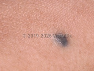
The blue nevus, also known as an acquired nevus, is a developmental lesion caused by aggregates of dendritic melanocytes that lie in the deep mucosa. The depth of the pigment creates a "Tyndall effect," which is what makes the melanin pigment appear blue. Although the literature reveals a 2:1 female predilection, this may be related to female patients seeking care more often and having more cosmetic concerns than male patients. It represents approximately one-fifth to one-fourth of all intraoral melanocytic nevi. Most nevi are noticed in the second to fourth decade of life and are usually asymptomatic.
Codes
ICD10CM:
D22.9 – Melanocytic nevi, unspecified
SNOMEDCT:
254806009 – Blue nevus of skin
D22.9 – Melanocytic nevi, unspecified
SNOMEDCT:
254806009 – Blue nevus of skin
Look For
Subscription Required
Diagnostic Pearls
Subscription Required
Differential Diagnosis & Pitfalls

To perform a comparison, select diagnoses from the classic differential
Subscription Required
Best Tests
Subscription Required
Management Pearls
Subscription Required
Therapy
Subscription Required
References
Subscription Required
Last Updated:12/06/2021
 Patient Information for Blue nevus - Oral Mucosal Lesion
Premium Feature
VisualDx Patient Handouts
Available in the Elite package
- Improve treatment compliance
- Reduce after-hours questions
- Increase patient engagement and satisfaction
- Written in clear, easy-to-understand language. No confusing jargon.
- Available in English and Spanish
- Print out or email directly to your patient
Upgrade Today

Blue nevus - Oral Mucosal Lesion
See also in: Overview,Hair and Scalp