Contents

SynopsisCodesLook ForDiagnostic PearlsDifferential Diagnosis & PitfallsBest TestsManagement PearlsTherapyDrug Reaction DataReferences

View all Images (36)

Other Resources UpToDate PubMed

Bursitis

Contributors: Juan Torres MD, Noah Craft MD, PhD
Other Resources UpToDate PubMed

Synopsis

Bursitis is swelling or enlargement of 1 or more of the > 150 bursae in the human body. Bursae are synovial pouches that reduce friction between adjacent tissues (eg, muscles, tendons, bony prominences). Bursitis may be acute or chronic in nature.

The location of bursae may be superficial (olecranon, patellar, infrapatellar, retrocalcaneal) or deep (anserine, subacromial, trochanteric, iliopsoas).

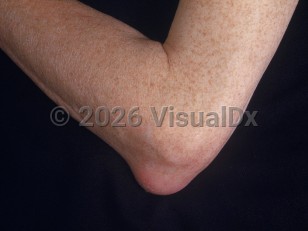
Commonly affected bursae are those over the extensor surfaces of the elbow (olecranon bursitis) and knee (prepatellar bursitis). Other commonly encountered bursitis sites are trochanteric bursitis near the greater trochanter of the femur and pes anserine bursitis over the anteromedial aspect of the tibia.

The etiology for bursitis is varied and can relate to the location of the bursitis. Generally bursitis is broken down into two broad categories: septic and nonseptic bursitis. Nonseptic bursitis may occur in the setting of inflammatory arthritis (eg, rheumatoid arthritis), trauma / injury, or prolonged localized pressure, or may be crystal induced (ie, gout or calcium pyrophosphate disease [CPPD]) or idiopathic. Shoulder bursitis may occur after routine vaccinations, often related to the position of the injection.

Septic bursitis usually results from trauma leading to direct inoculation of a bursa but may also be secondary to blunt trauma, a skin wound, or repetitive pressure applied over a joint. The source of infection is often from adjacent tissue, and overlying cellulitis may be associated with bursitis. Staphylococcus aureus is the most commonly isolated pathogen in septic bursitis.

Bursitis can occur at any age, but it is more commonly seen in adults and in the setting of overuse injuries. Repetitive activities (from sports or work) and certain occupations (laborer, gardener, mechanic, miner, and carpet layer) put one at greater risk for acquiring bursitis. Long-distance running is a risk factor for anserine bursitis. Trochanteric bursitis occurs more often in females than in males and has been associated with obesity, degenerative joint disease, and ipsilateral and contralateral hip arthritis. Certain health conditions such as gout, CPPD, and rheumatoid arthritis are predisposing factors, especially for olecranon and prepatellar bursitis. Risk factors for septic bursitis include having a suppressed immune system, such as those patients with malignancy, leukopenia, diabetes, renal failure, or recent use of systemic glucocorticoids.

Codes

ICD10CM:
M71.50 – Other bursitis, not elsewhere classified

SNOMEDCT:
84017003 – Bursitis

Look For

Subscription Required

Diagnostic Pearls

Subscription Required

Differential Diagnosis & Pitfalls

The most common diagnostic dilemma (particularly with acute inflammatory or septic bursitis) is differentiating bursitis from overlying soft tissue infection or from underlying septic arthritis. Many of the diagnoses on the list below can also be associated with a concurrent bursitis:
  • Septic arthritis (gonococcal or nongonococcal) – Systemic symptoms, painful joint, and joint aspirate with gram stain or cultures suggestive of microbial infection.
  • Lyme disease – Presence of erythema migrans and multiple risk factors (recent tick exposure in an endemic area during a high-risk season) may suggest Lyme etiology.
  • Cellulitis – Painful, warm, erythematous soft tissue infection with expanding border of involvement; signs of infection, fever.
  • Rheumatoid arthritis (RA) – Symmetric polyarthritis with active synovitis; may be seropositive (rheumatoid factor [RF], anticyclic citrullinated peptide [anti-CCP]) or seronegative.
  • Gout – Joint aspiration with crystal examination showing negative birefringent monosodium urate crystals with negative gram stain and culture.
  • Pseudogout (CPPD) – Joint aspiration revealing the presence of calcium pyrophosphate.
  • Osteoarthritis (OA) – Crepitation on exam; lack of erythema, warmth; symptoms worse with activity; typically involving weight-bearing joints and hands (especially base of the thumb); radiographs demonstrate degenerative change.
  • Spondyloarthritides (eg, psoriatic arthritis, reactive arthritis) – Demonstrates features of an inflammatory arthritis on exam; may present in several ways, including as an asymmetric monoarticular, oligoarticular, or polyarticular arthritis.
  • Epicondylitis – Commonly seen in lateral epicondylitis (pain with wrist extension) and medial epicondylitis (pain with wrist flexion) due to repetitive extension and flexion, respectively. Pain with palpation of the affected side of the epicondyle.
  • Enthesitis – Inflammation of where tendons, ligaments, or joint capsules attach to bone. Associated with spondyloarthropathy (psoriatic arthritis, ankylosing spondylitis, etc), periarticular bone erosion, and HLA-B27. Most commonly seen in lower limbs. Entheseal abnormalities can be detected by ultrasound.
  • Tendinosis – Associated with overuse injury. Frequently seen in aging athletes. Pain localized to the tendon due to microtearing of the tendon.
  • Iliotibial band syndrome (ITBS) – Distinguished from trochanteric bursitis by more inferior involvement along the lateral aspect of the thigh. Can also be associated with pain on the lateral epicondyle of the femur. This is seen largely in distance runners as an overuse injury. Look for a positive Noble test (pain produced on extension of leg from 90-degree flexion [patient is supine] while examiner applies pressure with thumb to the IT band). Note that ITBS can cause bursitis.

Best Tests

Subscription Required

Management Pearls

Subscription Required

Therapy

Subscription Required

Drug Reaction Data

Below is a list of drugs with literature evidence indicating an adverse association with this diagnosis. The list is continually updated through ongoing research and new medication approvals. Click on Citations to sort by number of citations or click on Medication to sort the medications alphabetically.

Subscription Required

References

Subscription Required

Last Updated:03/02/2021
Copyright © 2023 VisualDx®. All rights reserved.
Bursitis
Clinical image of Bursitis - imageId=2529215. Click to open in gallery.  caption: 'A faint pink nodule over the elbow.'
A faint pink nodule over the elbow.
Copyright © 2023 VisualDx®. All rights reserved.