Calcinosis cutis, or cutaneous calcification, is the deposition of insoluble calcium salts in the skin and subcutaneous tissue due to local dysregulation of calcium metabolism. Classically, disorders of calcium deposition can be broadly categorized into 5 main subtypes: dystrophic, metastatic, idiopathic, iatrogenic, and mixed.
In children, there is a male predilection with an earlier age of onset. Dystrophic and idiopathic calcification are observed more commonly than other types of calcium deposition. Dystrophic calcification usually arises in damaged skin in which local calcium metabolism is altered, allowing intracellular crystallization in the setting of normal serum calcium and phosphorus levels. Dystrophic calcification is typically seen in autoimmune connective tissue diseases, most commonly in juvenile dermatomyositis (DM) and the CREST form of systemic sclerosis. Approximately 50%-70% of children with juvenile DM will develop calcinosis cutis or some form of cutaneous calcification, in contrast to 10%-20% of patients with adult DM. The most frequently affected sites in DM are the elbows, knees, buttocks, and shoulders. In the CREST form of systemic sclerosis, the hands and upper extremities, often over bony prominences and tendons, are most commonly affected.
Other causes for dystrophic cutaneous calcification in children include cutaneous tumors and cysts such as pilomatricomas or pilar cysts, trauma (particularly associated with "heel sticks" in neonates or from other injection sites; see calcified nodules of the heel), infections (especially parasitic), and genetic disorders such as pseudoxanthoma elasticum and Ehlers-Danlos syndrome.
Subepidermal calcified nodules are idiopathic lesions that usually develop in children, although they can be seen in all age groups. They are hypothesized to occur as a result of trauma or calcification of a pre-existing milium, eccrine duct hamartoma, or hair follicle nevus.
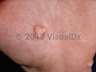
The most common clinical presentation is a solitary or multiple firm, jagged nodules. Lesions can extrude chalk-like calcium through the skin, which is often painful and may be associated with secondary infection.
Calcinosis universalis refers to the most severe form of dystrophic calcification. Usually observed in the setting of DM or other connective tissue diseases such as systemic lupus erythematosus, this variant involves extensive areas of cutaneous calcification in sheet-like masses and can cause severe functional impairment.
Calcinosis cutis in Infant/Neonate
Alerts and Notices
Important News & Links
Synopsis

Codes
ICD10CM:
L94.2 – Calcinosis cutis
SNOMEDCT:
21323007 – Calcinosis cutis
L94.2 – Calcinosis cutis
SNOMEDCT:
21323007 – Calcinosis cutis
Look For
Subscription Required
Diagnostic Pearls
Subscription Required
Differential Diagnosis & Pitfalls

To perform a comparison, select diagnoses from the classic differential
Subscription Required
Best Tests
Subscription Required
Management Pearls
Subscription Required
Therapy
Subscription Required
Drug Reaction Data
Subscription Required
References
Subscription Required
Last Reviewed:05/23/2020
Last Updated:06/01/2020
Last Updated:06/01/2020
Calcinosis cutis in Infant/Neonate