The melanocytic nevus is seen infrequently in the oral cavity, and presumably has a sequence of development similar to cutaneous melanocytic nevi.
The lesion can be identified at any age, and no sex predilection is seen.
Such lesions are asymptomatic, and no systemic signs, predisposing medical history, or risk factors are associated with the development of the intraoral melanocytic nevus.
The intraoral melanocytic nevus presumably evolves slowly, over a period of months to years.
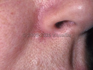
Common acquired nevus - Oral Mucosal Lesion
See also in: Overview,External and Internal Eye,Hair and ScalpAlerts and Notices
Important News & Links
Synopsis

Codes
ICD10CM:
D22.9 – Melanocytic nevi, unspecified
SNOMEDCT:
400096001 – Melanocytic nevus
D22.9 – Melanocytic nevi, unspecified
SNOMEDCT:
400096001 – Melanocytic nevus
Look For
Subscription Required
Diagnostic Pearls
Subscription Required
Differential Diagnosis & Pitfalls

To perform a comparison, select diagnoses from the classic differential
Subscription Required
Best Tests
Subscription Required
Management Pearls
Subscription Required
Therapy
Subscription Required
References
Subscription Required
Last Updated:02/25/2010
 Patient Information for Common acquired nevus - Oral Mucosal Lesion
Premium Feature
VisualDx Patient Handouts
Available in the Elite package
- Improve treatment compliance
- Reduce after-hours questions
- Increase patient engagement and satisfaction
- Written in clear, easy-to-understand language. No confusing jargon.
- Available in English and Spanish
- Print out or email directly to your patient
Upgrade Today

Common acquired nevus - Oral Mucosal Lesion
See also in: Overview,External and Internal Eye,Hair and Scalp