Contents

SynopsisCodesLook ForDiagnostic PearlsDifferential Diagnosis & PitfallsBest TestsManagement PearlsTherapyReferences

Information for Patients

View all Images (11)

Common acquired nevus - Hair and Scalp
See also in: Overview,External and Internal Eye,Oral Mucosal Lesion
Other Resources UpToDate PubMed

Common acquired nevus - Hair and Scalp

See also in: Overview,External and Internal Eye,Oral Mucosal Lesion
Contributors: William M. Lin MD, Sarah Hocker DO, Susan Burgin MD
Other Resources UpToDate PubMed

Synopsis

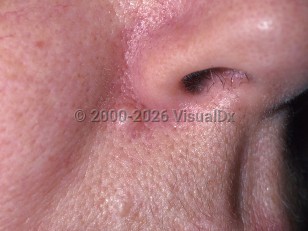
Common acquired nevi (moles) include junctional, dermal, and compound nevi, which are all considered benign. These distinctions are based upon the location of melanocytic nests in the epidermis, dermis, or both, respectively. Clinically, junctional nevi are flat (macular) whereas dermal and compound nevi are elevated relative to the surrounding skin (papular).

Nevi typically arise during childhood, adolescence, or very early adulthood and then senesce in later years. During pregnancy, existing nevi may darken and become noticeable to the patient. Compound nevi are more common in individuals with lighter skin phototypes; other forms of nevi (those on palms, soles, conjunctiva, and in the nail bed) are more common in individuals of African and Asian descent.

Codes

ICD10CM:
D22.9 – Melanocytic nevi, unspecified

SNOMEDCT:
400096001 – Melanocytic nevus

Look For

Subscription Required

Diagnostic Pearls

Subscription Required

Differential Diagnosis & Pitfalls

  • Melanoma
  • Atypical nevus
  • Acrochordon (skin tag)
  • Neurofibroma
  • Dermatofibroma
  • Blue nevus – typically macular, 2-5 mm, and with a blue hue
  • Combined nevus – two types of nevi that are seen together in one lesion; most commonly blue nevus and dermal or compound nevus
  • Recurrent melanocytic nevus – history of the initial biopsy is often critical for the dermatologist and/or pathologist. Recurrent nevi characteristically occur in women, 20-30 years of age, on the trunk within 6 months of the original biopsy. While many different clinical morphologies may be seen, it often manifests as a scar with variegated hyper- or hypopigmentation, linear streaking and halo, stippled and/or diffuse pigmentation patterns. While most cases do not pose a diagnostic challenge on histopathology, some specimens, especially partial biopsies, may look indistinguishable from melanoma on histopathologic grounds alone.
  • Desmoplastic nevus – typically small (< 1 cm) skin-colored to erythematous to mildly pigmented papule or nodule on the extremity of a young adult (average 30 years of age). Histopathology reveals spindled to epithelioid melanocytes within a fibrotic stroma.
  • Pigmented basal cell carcinoma
  • Wart

Best Tests

Subscription Required

Management Pearls

Subscription Required

Therapy

Subscription Required

References

Subscription Required

Last Updated:09/04/2018
Copyright © 2023 VisualDx®. All rights reserved.
Patient Information for Common acquired nevus - Hair and Scalp
Print E-Mail Images (11)
Contributors: Medical staff writer

Overview

A mole (nevus) is a non-cancerous (benign) skin lesion that is made up of the color-producing (pigment-producing) cells of the skin (melanocytes). A mole that is present at birth is referred to as a congenital nevus. A dysplastic nevus (discussed elsewhere) is a mole in which unusual (atypical) growth is noted. Moles (nevi, the plural of nevus) slowly enlarge evenly in all directions. After moles stop growing (stabilize), they may persist or they may become smaller (regress) later in life. Sun exposure and family tendency (heredity) play a role in the development of moles. Moles may sometimes become warm or red in color (inflamed) or irritated by friction from rubbing or contact with rough clothing or by other types of injury.

Who’s At Risk

Moles may occur in people of all races and ages, but they most commonly appear between the ages of 10 and 30. Individuals with light skin color are more likely to develop moles.

Signs & Symptoms

  • Moles may occur anywhere on the body, including the nails, palms, and soles.
  • Moles may be raised or flat.
  • Color of moles may vary from pink to skin colored to brown, but they may be darker in people with darker-skinned individuals.
  • Non-cancerous (benign) moles are usually alike on both sides (symmetrical), have smooth borders, have uniform color, and are generally smaller than the size of a pencil eraser (6 mm).

Self-Care Guidelines

  • Protective measures, such as avoiding skin exposure to sunlight during peak sun hours (10 AM to 3 PM), wearing protective clothing, and applying high-SPF sunscreen, are essential for reducing exposure to harmful ultraviolet (UV) light.
  • Monthly self-examination of the skin is helpful to detect new lesions or changes in existing lesions.
  • Make sure your moles are not signs of skin cancer (melanoma). Remember the ABCDEs of melanoma lesions:
    A - Asymmetry: One half of the lesion does not mirror the other half.
    B - Border: The borders are irregular or vague (indistinct).
    C - Color: More than one color may be noted within the mole.
    D - Diameter: Size greater than 6 mm (roughly the size of a pencil eraser) may be concerning.
    E - Evolving: Notable changes in the lesion over time are suspicious signs for skin cancer.

When to Seek Medical Care

  • The occurrence of a new mole in an adult over the age of 50 is unusual; if it occurs, see your doctor for evaluation.
  • People with multiple moles and unusual (atypical) moles should be examined by a dermatologist every 4–12 months, depending on their past history and family history.
  • It may be difficult to tell an atypical nevus from a normal nevus, so seek medical evaluation if you are unsure about the nature of a mole, if you note changes within a mole, or if a mole becomes irritated or painful.

Treatments

  • Non-cancerous (benign) moles do not require treatment, though they may be cosmetically removed.
  • If benign-appearing moles are inflamed or irritated, they can be surgically removed.

References


Freedberg, Irwin M., ed. Fitzpatrick's Dermatology in General Medicine. 6th ed. pp.889-893. New York: McGraw-Hill, 2003.
Copyright © 2023 VisualDx®. All rights reserved.
Common acquired nevus - Hair and Scalp
See also in: Overview,External and Internal Eye,Oral Mucosal Lesion
A medical illustration showing key findings of Common acquired nevus : Brown color, Pigmented papule, Smooth papule, Darkening skin lesion
Clinical image of Common acquired nevus - imageId=329792. Click to open in gallery.  caption: 'A skin-colored papule (intradermal nevus) at the nasal alar crease.'
A skin-colored papule (intradermal nevus) at the nasal alar crease.
Copyright © 2023 VisualDx®. All rights reserved.