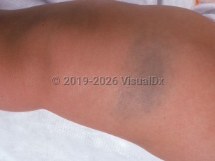
Contents

SynopsisCodesLook ForDiagnostic PearlsDifferential Diagnosis & PitfallsBest TestsManagement PearlsTherapyReferences

Information for Patients

View all Images (10)

Congenital dermal melanocytosis in Child
See also in: Anogenital
Other Resources UpToDate PubMed

Congenital dermal melanocytosis in Child

See also in: Anogenital
Contributors: William M. Lin MD, Craig N. Burkhart MD, Dean Morrell MD, Belinda Tan MD, PhD, Susan Burgin MD
Other Resources UpToDate PubMed

Synopsis

Congenital dermal melanocytosis (previously referred to as Mongolian spot) is a congenital, benign, blue to gray patch caused by an increased number of dermal melanocytes. These patches are commonly seen in the sacrococcygeal area in infants of Asian or African descent but may be found on any cutaneous surface in infants of all ethnicities. The pigmentation becomes most intense at 1 year of age, reaches its peak diameter by 2 years of age, and usually fades completely by adulthood.

Related topic: acquired dermal melanocytosis

Codes

ICD10CM:
Q82.8 – Other specified congenital malformations of skin

SNOMEDCT:
40467008 – Mongolian spot

Look For

Subscription Required

Diagnostic Pearls

Subscription Required

Differential Diagnosis & Pitfalls

  • Child physical abuse – Childhood abuse affects children of all ages and economic and cultural backgrounds. It can often be challenging to differentiate findings attributable to child abuse from benign anogenital skin conditions. Abuse can present with purpura and ecchymoses: changes color from blue to green to brown over days to weeks, uneven pigmentation, tender to palpation. Documentation of congenital dermal melanocytosis may be helpful in avoiding any confusion.
  • Congenital melanocytic nevus – Brown, well-defined, surface change (eg, papular lesions).
  • Blue nevus – Usually much smaller, typically no more than 5 mm in diameter, well-defined.
  • Nevus of Ito – Often speckled, progresses with age, located over the shoulder, clavicle, and scapular region.
  • Nevus of Ota – Often speckled, progresses with age, located in the periorbital region.
  • Venous malformation – Usually palpable, may swell or change in coloration when dependent or crying.
  • Infantile hemangioma – Overlying telangiectasias, may swell or deepen in coloration when dependent or crying, rapidly elevates over days to weeks.

Best Tests

Subscription Required

Management Pearls

Subscription Required

Therapy

Subscription Required

References

Subscription Required

Last Updated:02/08/2021
Copyright © 2023 VisualDx®. All rights reserved.
Patient Information for Congenital dermal melanocytosis in Child
Print E-Mail Images (10)
Contributors: Medical staff writer

Overview

Blue-gray spots, commonly referred to as congenital dermal melanocytosis, are large flat lesions that are usually found on the lower back or buttocks of infants at birth. They can occasionally be found on the legs or shoulders of infants, but this is less common. The color of blue-gray spots ranges from deep brown to slate gray or blue-black. They are caused by collections of pigment-producing cells (melanocytes) located in a deeper layer in the skin and are the most common type of birthmark.

Who’s At Risk

Blue-gray spots are seen in approximately 90% of African Americans and Native Americans, roughly 80% of patients of Asian descent, 70% of Hispanic patients, and between 5%-10% of white patients. Generally, male and female infants are equally affected, although some studies suggest a slightly higher incidence of these birthmarks in males. Additionally, full-term infants are more likely to have blue-gray spots than preterm infants.

Signs & Symptoms

Blue-gray spots may be single or multiple, and they range in size from a few millimeters to 10 centimeters or more in diameter. They are present at birth and, due to their appearance, can be confused for a bruise.

Self-Care Guidelines

There is no self-care for blue-gray spots, though they naturally fade within the first years of life. In very rare cases, they persist into school age years and even into adulthood.

When to Seek Medical Care

Once the diagnosis of a blue-gray spot is made, there is essentially nothing to do except observe the spot over time and wait for resolution. If the spot does not fade within the first years of life, see your child's physician. Additionally, because this birthmark appears similarly to bruises, if you have any questions about whether or not it was present at birth, it is best to seek medical care.

Treatments

There is no therapy or treatment for this diagnosis in infants because it is a harmless condition that should resolve on its own. However, large and numerous spots that persist after a year of life may be associated with rare genetic disorders in a very small number of cases. If there is a strong family history of genetic disorders along with this condition, the child's physician may decide to send blood to the lab to test for specific genetic diseases, which each have their own specific treatments.

References


Ashrafti MR, Shabanian R, Mohammadi M, Kavusi S. Extensive Mongolian Spots: a clinical sign merits special attention. Pediatr Neurol. 2006;34(2):143-145.

Behrman RE, Kliegman R, Jensen HB, eds. Nelson Textbook of Pediatrics. 18th ed. Philadelphia, PA: WB Saunders; 2007.

Paller AS, Mancini AJ. Hurwitz Clinical Pediatric Dermatology: A Textbook of Skin Disorders of Childhood and Adolescence. Philadelphia, PA: Elsevier; 2005.
Copyright © 2023 VisualDx®. All rights reserved.
Congenital dermal melanocytosis in Child
See also in: Anogenital
A medical illustration showing key findings of Congenital dermal melanocytosis : Blue color, Hyperpigmented patch
Clinical image of Congenital dermal melanocytosis - imageId=779962. Click to open in gallery.  caption: 'A large blue-gray patch on the thigh.'
A large blue-gray patch on the thigh.
Copyright © 2023 VisualDx®. All rights reserved.