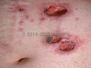
Pathogenic forms of Entamoeba histolytica cause cutaneous amebiasis, which usually results from direct extension of intestinal amebiasis. Involvement of the female genitals occurs by extension of rectal amebiasis. Colonic surgery on the involved amebic colon, hepatic amebiasis, and direct inoculation are also routes for skin involvement.
In general, cutaneous disease commonly presents as one or more painful ulcers that can become necrotic. Notably, ulcers tend to enlarge rapidly. If left untreated, progression is rapid and massive destruction of skin and subcutaneous tissues may ensue.
Cutaneous amebiasis - Anogenital in
See also in: OverviewAlerts and Notices
Important News & Links
Synopsis

Codes
ICD10CM:
A06.7 – Cutaneous amebiasis
SNOMEDCT:
111910009 – Amebiasis
A06.7 – Cutaneous amebiasis
SNOMEDCT:
111910009 – Amebiasis
Look For
Subscription Required
Diagnostic Pearls
Subscription Required
Differential Diagnosis & Pitfalls

To perform a comparison, select diagnoses from the classic differential
Subscription Required
Best Tests
Subscription Required
Management Pearls
Subscription Required
Therapy
Subscription Required
References
Subscription Required
Last Reviewed:03/20/2017
Last Updated:03/20/2017
Last Updated:03/20/2017
Cutaneous amebiasis - Anogenital in
See also in: Overview