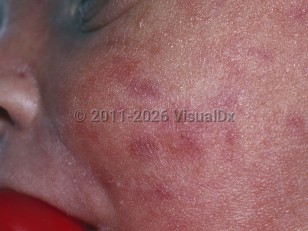
Cutaneous extramedullary hematopoiesis in Adult
Alerts and Notices
Important News & Links
Synopsis

Cutaneous extramedullary hematopoiesis is a rare cutaneous manifestation of myelofibrosis, where hematopoiesis occurs in the skin rather than the bone marrow, causing polymorphic skin lesions. The condition most often presents as bilateral lower extremity ulcers, but it may also cause papules, nodules, and plaques on any body part. These lesions typically begin as pink erythematous lesions, which become violaceous, hemorrhagic, and ulcerated over time. In addition, the lesions also tend to increase in number and size if the underlying hematological condition remains untreated.
(Of note, cutaneous extramedullary hematopoiesis is a normal occurrence during early embryogenesis, and it occurs until about the fifth month of gestation. Cutaneous extramedullary hematopoiesis in pre-term or full-term neonates is thought to be an accentuation of a normal physiologic process. These lesions resolve spontaneously 3-4 weeks after birth without intervention. Multiple reports suggest also that cutaneous extramedullary hematopoiesis in neonates may be precipitated by congenital infections, erythroblastosis fetalis, and twin transfusion syndrome.)
(Of note, cutaneous extramedullary hematopoiesis is a normal occurrence during early embryogenesis, and it occurs until about the fifth month of gestation. Cutaneous extramedullary hematopoiesis in pre-term or full-term neonates is thought to be an accentuation of a normal physiologic process. These lesions resolve spontaneously 3-4 weeks after birth without intervention. Multiple reports suggest also that cutaneous extramedullary hematopoiesis in neonates may be precipitated by congenital infections, erythroblastosis fetalis, and twin transfusion syndrome.)
Codes
ICD10CM:
D75.9 – Disease of blood and blood-forming organs, unspecified
SNOMEDCT:
73241006 – Abnormal hematopoiesis
D75.9 – Disease of blood and blood-forming organs, unspecified
SNOMEDCT:
73241006 – Abnormal hematopoiesis
Look For
Subscription Required
Diagnostic Pearls
Subscription Required
Differential Diagnosis & Pitfalls

To perform a comparison, select diagnoses from the classic differential
Subscription Required
Best Tests
Subscription Required
Management Pearls
Subscription Required
Therapy
Subscription Required
References
Subscription Required
Last Updated:12/19/2017
Cutaneous extramedullary hematopoiesis in Adult

