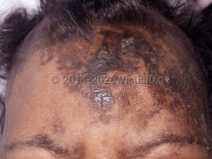
Dermatosis neglecta (DN) is a condition characterized by adherent scales that form due to inadequate cleaning or exfoliation of the skin. Whether due to deliberately poor or unwittingly neglectful personal hygiene, the lack of washing over the course of months allows for the accumulation of sebum, sweat, keratin, debris, bacteria, and yeast on unwashed areas of the skin. Pruritus may develop over the site, but there are no systemic symptoms. Washing with soap, water, and a washcloth for friction or with isopropyl alcohol-soaked gauze will often result in complete removal of the lesions.
DN may present in individuals with an underlying physical disability, hyperesthesia, previous trauma, or with a psychiatric etiology, resulting in areas of skin that remain persistently unwashed. Although not a life-threatening or contagious condition, it can be cosmetically bothersome.
Dermatosis neglecta in Adult
Alerts and Notices
Important News & Links
Synopsis

Codes
ICD10CM:
L85.9 – Epidermal thickening, unspecified
SNOMEDCT:
402343006 – Retention hyperkeratosis
L85.9 – Epidermal thickening, unspecified
SNOMEDCT:
402343006 – Retention hyperkeratosis
Look For
Subscription Required
Diagnostic Pearls
Subscription Required
Differential Diagnosis & Pitfalls

To perform a comparison, select diagnoses from the classic differential
Subscription Required
Best Tests
Subscription Required
Management Pearls
Subscription Required
Therapy
Subscription Required
References
Subscription Required
Last Reviewed:03/01/2021
Last Updated:03/01/2021
Last Updated:03/01/2021
Dermatosis neglecta in Adult