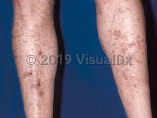
Diabetic dermopathy, commonly known as shin spots, is the most common cutaneous finding in patients with diabetes mellitus, presenting in up to 50% of diabetic patients. The etiology of diabetic dermopathy is unclear. Previously, ischemia had been thought to be a causative factor, but studies have found that blood flow to the dermopathy lesions was actually increased compared with other surrounding areas. Trauma is thought to be a causative factor.
There is no clear variation of incidence between diabetic dermopathy in patients with noninsulin-dependent diabetes mellitus versus those with insulin-dependent diabetes mellitus. There is, however, a correlation between the presence of skin lesions and the presence of microangiopathic complications (retinal, neuropathic, and/or nephrogenic). Diabetic dermopathy appears to be a useful clinical finding for detection of these diabetic complications.
The incidence of diabetic dermopathy increases with age. It is typically seen in patients aged older than 50. Men show an increased incidence compared to women.
Although located bilaterally, the distribution of lesions is asymmetric. Lesions do not itch or cause pain. Poor long-term blood sugar control, which increases risk of diabetic microangiopathic complications, is seen in diabetic dermopathy. There is no correlation between diabetic dermopathy and obesity or hypertension.
Related topics: diabetes mellitus type 1, diabetes mellitus type 2
Diabetic dermopathy
Alerts and Notices
Important News & Links
Synopsis

Codes
ICD10CM:
E13.620 – Other specified diabetes mellitus with diabetic dermatitis
SNOMEDCT:
238982009 – Dermopathy due to diabetes mellitus
E13.620 – Other specified diabetes mellitus with diabetic dermatitis
SNOMEDCT:
238982009 – Dermopathy due to diabetes mellitus
Look For
Subscription Required
Diagnostic Pearls
Subscription Required
Differential Diagnosis & Pitfalls

To perform a comparison, select diagnoses from the classic differential
Subscription Required
Best Tests
Subscription Required
Management Pearls
Subscription Required
Therapy
Subscription Required
References
Subscription Required
Last Reviewed:10/28/2019
Last Updated:03/12/2024
Last Updated:03/12/2024
 Patient Information for Diabetic dermopathy
Premium Feature
VisualDx Patient Handouts
Available in the Elite package
- Improve treatment compliance
- Reduce after-hours questions
- Increase patient engagement and satisfaction
- Written in clear, easy-to-understand language. No confusing jargon.
- Available in English and Spanish
- Print out or email directly to your patient
Upgrade Today

Diabetic dermopathy