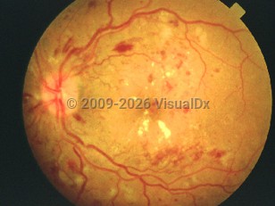
Diabetic retinopathy is the most common cause of blindness in adults between the ages of 20 and 64. It can be classified as nonproliferative (NPDR) or proliferative (PDR). In individuals with chronically elevated blood sugars, the retinal blood vessels undergo biochemical and physiologic changes that damage the vascular endothelium. Early in the nonproliferative state of the disease, microaneurysms develop in the weakened vessel walls. With increased vascular permeability, retinal edema and hemorrhages occur. As the disease progresses, the retina tissue becomes ischemic and new abnormal blood vessels form. When these abnormal vessels gain entry outside of the retina into the posterior hyaloid, the term PDR is used to describe the disease.
Patients with diabetic retinopathy are often asymptomatic. As the disease progresses, they may sense vision loss due to macular edema, hemorrhage, or retinal detachment. The risk of diabetic retinopathy increases with the duration of diabetes and the patient's age. Nearly 99% of individuals with type 1 diabetes and 60% of individuals with type 2 diabetes develop diabetic retinopathy after 20 years of the diagnosis. The condition is more common in individuals of African and Mexican descent compared with those of Northern European descent. Cardiovascular disease risk factors, such as hypertension, are also associated with advanced diabetic retinopathy.
Pediatric Patient Considerations:
Diabetic retinopathy is rarely found in children younger than 10. The risk of retinopathy developing in children with diabetes increases after puberty.
Related topics: diabetes mellitus type 1, diabetes mellitus type 2
Diabetic retinopathy - External and Internal Eye
Alerts and Notices
Important News & Links
Synopsis

Codes
ICD10CM:
E10.311 – Type 1 diabetes mellitus with unspecified diabetic retinopathy with macular edema
E10.3299 – Type 1 diabetes mellitus with mild nonproliferative diabetic retinopathy without macular edema, unspecified eye
E11.311 – Type 2 diabetes mellitus with unspecified diabetic retinopathy with macular edema
SNOMEDCT:
4855003 – Diabetic retinopathy
Look For
Subscription Required
Diagnostic Pearls
Subscription Required
Differential Diagnosis & Pitfalls

To perform a comparison, select diagnoses from the classic differential
Subscription Required
Best Tests
Subscription Required
Management Pearls
Subscription Required
Therapy
Subscription Required
References
Subscription Required
Last Reviewed:03/09/2017
Last Updated:08/06/2023
Last Updated:08/06/2023
Diabetic retinopathy - External and Internal Eye