Dissecting cellulitis of the scalp, also known as perifolliculitis capitis abscedens et suffodiens (PCAS), is a neutrophilic scarring alopecia with an abnormal inflammatory response to staphylococcal antigens. This association is linked to a propensity for follicular occlusion and dramatic secondary inflammatory changes to proinflammatory stimuli, such as bacterial infection. The follicle occludes, dilates, and ruptures, and the keratin promotes an inflammatory response in conjunction with a secondary staphylococcal infection attracting neutrophils.
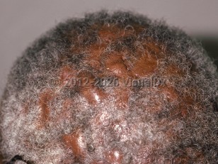
The condition typically affects Black men between the ages of 20 and 40, but it can occur in other races and ethnicities, in women, and in children. Dissecting cellulitis usually affects the vertex (although the entire scalp can be involved), producing boggy or fluctuant pustules and nodules. Patients complain of pain and of a disfiguring appearance. If a nodule is pressed, pus or serosanguineous fluid may extrude from one or more of the orifices.
The disease may wax and wane over several years, later producing dermal fibrosis, sinus tracts, and hypertrophic scarring with alopecia. There is an increased risk of squamous cell carcinoma in patients with long-standing disease.
Dissecting cellulitis may be isolated or may occur as part of a follicular occlusion triad that includes acne conglobata and hidradenitis suppurativa or a tetrad that additionally includes pilonidal cysts.
It has been rarely associated with skull osteomyelitis, arthritis with keratitis, pyoderma vegetans, pityriasis rubra pilaris, keratitis-ichthyosis-deafness syndrome, Crohn disease, and pyoderma gangrenosum.
In contrast to classic cellulitis, dissecting cellulitis of the scalp demonstrates prominent nodularity. Lesions may be fluctuant and/or draining. Location and patient demographic factors, such as race, are important diagnostic clues.
Dissecting cellulitis of scalp - Cellulitis DDx
See also in: Hair and ScalpAlerts and Notices
Important News & Links
Synopsis

Codes
ICD10CM:
L66.3 – Perifolliculitis capitis abscedens
SNOMEDCT:
77333008 – Dissecting cellulitis of scalp
L66.3 – Perifolliculitis capitis abscedens
SNOMEDCT:
77333008 – Dissecting cellulitis of scalp
Look For
Subscription Required
Diagnostic Pearls
Subscription Required
Differential Diagnosis & Pitfalls

To perform a comparison, select diagnoses from the classic differential
Subscription Required
Best Tests
Subscription Required
Management Pearls
Subscription Required
Therapy
Subscription Required
References
Subscription Required
Last Reviewed:02/06/2020
Last Updated:05/18/2023
Last Updated:05/18/2023
 Patient Information for Dissecting cellulitis of scalp - Cellulitis DDx
Premium Feature
VisualDx Patient Handouts
Available in the Elite package
- Improve treatment compliance
- Reduce after-hours questions
- Increase patient engagement and satisfaction
- Written in clear, easy-to-understand language. No confusing jargon.
- Available in English and Spanish
- Print out or email directly to your patient
Upgrade Today

Dissecting cellulitis of scalp - Cellulitis DDx
See also in: Hair and Scalp