Cephalocele is a malformation caused by intracranial structures herniating through an opening in the skull. Encephaloceles specifically contain both meninges and brain, while meningoceles contain meninges and cerebrospinal fluid (CSF).
When the neural tube does not close properly during development, this can result in an encephalocele.
Cephaloceles are quite rare but can occur more frequently in patients with a family history of cephaloceles.
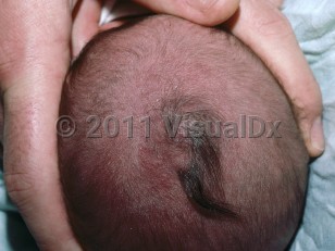
Encephaloceles frequently occur as soft nodules or masses that grow as the infant grows and grow acutely with crying or during a Valsalva maneuver. The most common locations for encephaloceles are the occiput and the vertex located near the midline of the skull. However, encephaloceles have been reported near the nasal glabella and near the pharyngeal region.
Encephaloceles commonly lead to early deformities and can be mistaken for deeply seated hemangiomas or vascular malformations. The deformity can lead to the misperception of hypertelorism as well.
Repetitive cephaloceles in families may be attributed to specific genes responsible for controlling individual closure sites. Patients may report a history of meningitis.
Related topic: atretic encephalocele
Encephalocele
Alerts and Notices
Important News & Links
Synopsis

Codes
ICD10CM:
Q01.9 – Encephalocele, unspecified
SNOMEDCT:
55999004 – Encephalocele
Q01.9 – Encephalocele, unspecified
SNOMEDCT:
55999004 – Encephalocele
Look For
Subscription Required
Diagnostic Pearls
Subscription Required
Differential Diagnosis & Pitfalls

To perform a comparison, select diagnoses from the classic differential
Subscription Required
Best Tests
Subscription Required
Management Pearls
Subscription Required
Therapy
Subscription Required
References
Subscription Required
Last Updated:10/30/2022
Encephalocele