Causes / typical injury mechanism: Enchondroma is a benign, hyaline-cartilage producing tumor located in the medullary canal of the diaphysis or metaphysis of long bones.
Ollier disease and Maffucci syndrome are nonhereditary conditions that present as multiple enchondromas. Ollier disease involves multiple asymmetric enchondromas of the digits. It is most commonly seen in childhood. These slow-growing tumors usually stop growing after puberty, but masses can create deformity and limb asymmetry. Pathologic fractures may be seen. Ollier disease has a 15%-20% risk for secondary chondrosarcoma.
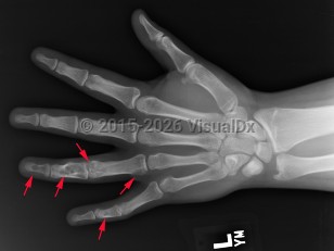
Classic history and presentation: The proximal phalanx is the typical location. These tumors are also common incidental findings in the distal femur, proximal humerus, and tibia. Mean length is usually less than 5 cm.
Solitary enchondroma itself has about a 1% risk of malignant transformation into chondrosarcoma. It is important to distinguish enchondroma from chondrosarcoma, a malignant tumor. Patients with chondrosarcoma of the extremity are more likely to present with nighttime pain, pathologic fracture, endosteal cortical scalloping, bony reactive changes, and larger lesions (> 6 cm). Assume any pelvic cartilage tumor in an adult is a chondrosarcoma until proven otherwise. Rib and scapula lesions are also more likely to be chondrosarcomas.
Prevalence: Enchondroma is a latent lesion that comprises 15%-25% of cartilage tumors. It is the most common hand tumor, accounting for 90% of cases. Enchondroma usually presents in the third and fourth decades of life and occurs equally between both sexes.
Pathophysiology: Enchondroma pathophysiology is believed to involve incomplete endochondral ossification in which physeal remnants become entrapped in the medullary cavity of the metaphysis and proliferate. In the hand, enchondromas are typically diagnosed after a pathologic fracture. Elsewhere, they are typically an asymptomatic incidental finding on x-ray or advanced imaging performed for other reasons. Pain is usually due to a nearby abnormality rather than the tumor itself.
Enchondroma in Adult
Alerts and Notices
Important News & Links
Synopsis
Codes
ICD10CM:
D16.10 – Benign neoplasm of short bones of unspecified upper limb
SNOMEDCT:
423699002 – Enchondroma
D16.10 – Benign neoplasm of short bones of unspecified upper limb
SNOMEDCT:
423699002 – Enchondroma
Look For
Subscription Required
Diagnostic Pearls
Subscription Required
Differential Diagnosis & Pitfalls

To perform a comparison, select diagnoses from the classic differential
Subscription Required
Best Tests
Subscription Required
Management Pearls
Subscription Required
Therapy
Subscription Required
References
Subscription Required
Last Reviewed:08/12/2020
Last Updated:04/14/2024
Last Updated:04/14/2024
Enchondroma in Adult