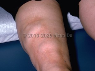
Classically, EF presents abruptly with symmetrical erythema and edema of the affected extremities sparing the hands, feet, and face. This progresses rapidly to induration and fibrosis, which is usually accompanied by pain and joint contractures, resulting in a decreased range of motion and paresthesias. The skin in EF is often bound down, accentuating the natural planes between muscles and vasculature, and revealing linear depressions along superficial veins, causing the "groove sign." This clinical sign, along with peau d'orange changes, seem to be less common in the pediatric age group, whereas the articular manifestations, including acral tendon retractions resulting in the "prayer sign," are nearly universal in this population.
The etiology is unknown, and while EF is especially rare in children, it can and has been noted to occur in children as young as 1 year. A younger age of onset is associated with a greater risk of progression to cutaneous fibrosis and joint contractures. White individuals are most often reported to have this condition. Age of onset has a bimodal distribution, with pediatric disease occurring in childhood to adolescence, with studies reporting a mean age of onset between 8 and 13 years. There is a female preponderance in pediatric EF. The adult form develops between ages 30 and 50 years.
The etiology is unknown, and while EF is especially rare in children, it can and has been noted to occur in children as young as 1 year. A younger age of onset is associated with a greater risk of progression to cutaneous fibrosis and joint contractures. White individuals are most often reported to have this condition. Age of onset has a bimodal distribution, with pediatric disease occurring in childhood to adolescence, with studies reporting a mean age of onset between 8 and 13 years. There is a female preponderance in pediatric EF. The adult form develops between ages 30 and 50 years.
A history of strenuous physical activity preceding the clinical findings of EF occurs in approximately 30% of patients, although this is less noted in the pediatric population. In pediatric EF, nonspecific infections are more often a trigger than medications, as antihypertensives and statins are less commonly used in this age group. While concurrent autoimmune conditions and neoplasia seem to be less common in pediatric EF versus adult-onset disease, associated hypergammaglobulinemia is much more frequent.
Trunk involvement and the presence of peau d'orange have been associated with a poorer prognosis. Features that distinguish EF from deep morphea and scleroderma are a peripheral eosinophilia (in 60%-80% of patients), hypergammaglobulinemia (in 20%-70% of patients), and an absence of Raynaud phenomenon that is common to systemic sclerosis. In a retrospective study, 21 of 60 patients (35%) had concurrent plaque morphea. Antinuclear antibody (ANA) titers are normal in EF and the ESR is usually elevated.
Trunk involvement and the presence of peau d'orange have been associated with a poorer prognosis. Features that distinguish EF from deep morphea and scleroderma are a peripheral eosinophilia (in 60%-80% of patients), hypergammaglobulinemia (in 20%-70% of patients), and an absence of Raynaud phenomenon that is common to systemic sclerosis. In a retrospective study, 21 of 60 patients (35%) had concurrent plaque morphea. Antinuclear antibody (ANA) titers are normal in EF and the ESR is usually elevated.