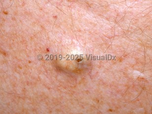
An epidermoid cyst, also known as an epidermal inclusion cyst or a sebaceous cyst, is a semisolid cyst. The cyst wall is stratified squamous epithelium, and the contents consist of macerated keratin and lipid-rich debris. Epidermoid cysts are commonly occurring lesions that can arise on the scalp, face, trunk, extremities, in the mouth, or on the genitals at any age, although they are most common in adults. They are also more common in men.
Several etiologic factors have been implicated in the formation of epidermoid cysts, including traumatic or iatrogenic implantation of epidermal elements, sequestration of epidermal rests, occlusion of the eccrine duct or pilosebaceous unit, and human papillomavirus (HPV) infection. Smoking may contribute to the development of multiple epidermoid cysts on the face. Epidermoid cysts are a feature of several hereditary syndromes, such as Gardner syndrome, pachyonychia congenita, and the basal cell nevus syndrome.
Epidermoid cysts are benign and usually asymptomatic, but they may be painful if ruptured or infected. Rarely, malignancies such as basal cell carcinoma, squamous cell carcinoma, and mycosis fungoides have developed within these cysts.
Pediatric Patient Considerations:
It is rare to see an epidermoid cyst in a prepubertal patient; in such cases, other diagnoses should be carefully considered.
Epidermoid cyst - Hair and Scalp
See also in: Overview,AnogenitalAlerts and Notices
Important News & Links
Synopsis

Codes
ICD10CM:
L72.0 – Epidermal cyst
SNOMEDCT:
419893006 – Epidermoid cyst
L72.0 – Epidermal cyst
SNOMEDCT:
419893006 – Epidermoid cyst
Look For
Subscription Required
Diagnostic Pearls
Subscription Required
Differential Diagnosis & Pitfalls

To perform a comparison, select diagnoses from the classic differential
Subscription Required
Best Tests
Subscription Required
Management Pearls
Subscription Required
Therapy
Subscription Required
References
Subscription Required
Last Reviewed:05/26/2024
Last Updated:05/27/2024
Last Updated:05/27/2024
 Patient Information for Epidermoid cyst - Hair and Scalp
Premium Feature
VisualDx Patient Handouts
Available in the Elite package
- Improve treatment compliance
- Reduce after-hours questions
- Increase patient engagement and satisfaction
- Written in clear, easy-to-understand language. No confusing jargon.
- Available in English and Spanish
- Print out or email directly to your patient
Upgrade Today

Epidermoid cyst - Hair and Scalp
See also in: Overview,Anogenital