An intracranial epidural hematoma (EDH) occurs when blood accumulates between the dura and inner table of the calvarium. The classic etiology of the bleed is traumatic injury to the middle meningeal artery following a skull fracture. The source of bleeding is arterial in 85% of cases; in 15% of cases, the etiology is venous from a meningeal vein or dural venous sinus injury. As blood accumulates in the space between the outer layer of dura and skull, it strips the dura and forms a biconvex (lenticular or football-shaped) hematoma. The dura is very adherent to the overlying cranial sutures, so the hematoma is often limited. It is more commonly seen in children and adults, whose dura is less adherent to the inner table of the skull, as opposed to infants < 2 years of age or the elderly.
The classic presentation involves a brief post-traumatic loss of consciousness, then several minutes to hours of a lucid interval, followed by obtundation and focal neurologic deficits. While this classic clinical description is widely taught, it is seen in < 20% of cases. Other less specific signs and symptoms include headache, nausea and emesis, seizures, neurologic deficits (contralateral weakness, hyperreflexia), papilledema, pupil-involving third-nerve palsy, somnolence, or coma.
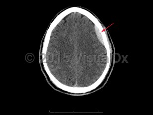
Epidural intracranial hematoma
Alerts and Notices
Important News & Links
Synopsis

Codes
ICD10CM:
S06.4X0A – Epidural hemorrhage without loss of consciousness, initial encounter
S06.4X0S – Epidural hemorrhage without loss of consciousness, sequela
S06.4X9A – Epidural hemorrhage with loss of consciousness of unspecified duration, initial encounter
SNOMEDCT:
428268007 – Extradural intracranial hematoma
Look For
Subscription Required
Diagnostic Pearls
Subscription Required
Differential Diagnosis & Pitfalls

To perform a comparison, select diagnoses from the classic differential
Subscription Required
Best Tests
Subscription Required
Management Pearls
Subscription Required
Therapy
Subscription Required
Drug Reaction Data
Subscription Required
References
Subscription Required
Last Reviewed:08/22/2017
Last Updated:08/30/2017
Last Updated:08/30/2017
Epidural intracranial hematoma