Contents

SynopsisCodesLook ForDiagnostic PearlsDifferential Diagnosis & PitfallsBest TestsManagement PearlsTherapyDrug Reaction DataReferences

Information for Patients

View all Images (31)

Erythema multiforme - Oral Mucosal Lesion
See also in: Overview,Anogenital
Other Resources UpToDate PubMed

Erythema multiforme - Oral Mucosal Lesion

See also in: Overview,Anogenital
Contributors: Erin X. Wei MD, Susan Burgin MD, Belinda Tan MD, PhD, Carl Allen DDS, MSD, Sook-Bin Woo MS, DMD, MMSc
Other Resources UpToDate PubMed

Synopsis

Erythema multiforme (EM) is a self-limited hypersensitivity reaction of the skin and mucous membranes characterized by the acute onset of fixed lesions of concentric color change (target lesions). Two subtypes exist: EM major and EM minor. Key differences between the EM subtypes include mucosal involvement and systemic symptoms such as fever, arthralgias, and asthenias seen in the major subtype. Prodromal symptoms occasionally can be associated.

Recurrent EM occurs in a subset of patients and has been variably defined as more than 1, more than 2, or more than 6 flares per year.

Persistent EM is uncommon and refers to chronic, continuous presence of EM or outbreaks separated by 15 days or less.

In adults, the primary trigger for EM is herpes simplex virus (HSV), which is estimated to incite about 90% of cases. EM has been reported with other infections including histoplasmosis, Epstein-Barr virus, and, most recently, COVID-19. Medication can also be a trigger. In children, important additional triggers to consider include drugs (particularly penicillin), group A Streptococcus, and Epstein-Barr virus, among other viruses and bacteria. Idiopathic cases have also been seen. 

Typically, all cutaneous lesions appear within 24-72 hours and persist for 1-4 weeks before fading. The eruption recurs on repeated exposure to the inciting agent. 

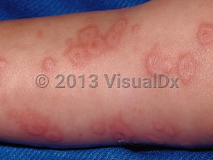
EM demonstrates classical target lesions, raised atypical target lesions, or both concomitantly on the skin.

The following points should be kept in mind when a diagnosis of EM is being considered:
  • Herpes labialis may typically precede development of EM but may sometimes develop concomitantly or manifest after the onset of EM (In almost half of all cases, herpes labialis precedes EM.)
  • Classical target lesions are well-defined circular lesions that are less than 3 cm in diameter and have 3 distinct color zones and a central zone that has a bulla or crust.
  • Atypical target lesions are palpable, poorly defined, circular lesions that have 2 distinct color zones. Raised atypical targets are a subtype of atypical targets that have a vesicle or bulla centrally.
  • EM is not considered within the same disease spectrum as Stevens-Johnson syndrome (SJS) / toxic epidermal necrolysis (TEN) and confers no risk of progressing to TEN.
  • EM major refers to the presence of significant mucosal involvement in a case of EM, whereas in EM minor, mucosal involvement is absent or minimal.
  • In EM major, painful intraoral and lip erosions eventuate from vesicles or bullae. Secondary crusting, including hemorrhagic crusting, then develops.

Codes

ICD10CM:
L51.9 – Erythema multiforme, unspecified

SNOMEDCT:
36715001 – Erythema multiforme

Look For

Subscription Required

Diagnostic Pearls

Subscription Required

Differential Diagnosis & Pitfalls

To perform a comparison, select diagnoses from the classic differential

Subscription Required

Best Tests

Subscription Required

Management Pearls

Subscription Required

Therapy

Subscription Required

Drug Reaction Data

Subscription Required

References

Subscription Required

Last Reviewed:12/19/2022
Last Updated:02/11/2023
Copyright © 2024 VisualDx®. All rights reserved.
Patient Information for Erythema multiforme - Oral Mucosal Lesion
Print E-Mail Images (31)
Contributors: Medical staff writer
Premium Feature
VisualDx Patient Handouts
Available in the Elite package
  • Improve treatment compliance
  • Reduce after-hours questions
  • Increase patient engagement and satisfaction
  • Written in clear, easy-to-understand language. No confusing jargon.
  • Available in English and Spanish
  • Print out or email directly to your patient
Copyright © 2024 VisualDx®. All rights reserved.
Erythema multiforme - Oral Mucosal Lesion
See also in: Overview,Anogenital
A medical illustration showing key findings of Erythema multiforme (Skin) : Scattered many
Clinical image of Erythema multiforme - imageId=30005. Click to open in gallery.  caption: 'Edematous and erythematous papules and plaques, some with a target-like appearance on the leg.'
Edematous and erythematous papules and plaques, some with a target-like appearance on the leg.
Copyright © 2024 VisualDx®. All rights reserved.