Contents

SynopsisCodesLook ForDiagnostic PearlsDifferential Diagnosis & PitfallsBest TestsManagement PearlsTherapyDrug Reaction DataReferences

Information for Patients

View all Images (4)

Follicular conjunctivitis - External and Internal Eye
Other Resources UpToDate PubMed

Follicular conjunctivitis - External and Internal Eye

Contributors: Brandon D. Ayres MD, Christopher Rapuano MD, Harvey A. Brown MD, Sunir J. Garg MD, Lauren Patty Daskivich MD, MSHS
Other Resources UpToDate PubMed

Synopsis

Most forms of follicular conjunctivitis are a local host response to an exogenous substance or agent producing prominent subconjunctival lymphoid follicles. Viruses, chlamydia, and bacteria can produce the problem. Careful history, clinical examination, and simple lab tests are required to ferret out specific causes. Symptoms and signs depend on the causative agent but may include watery discharge, conjunctival hyperemia, sandy/gravely feeling, blurry vision, and photophobia. There are both acute and chronic forms of follicular conjunctivitis with the latter defined as those cases of follicular conjunctivitis that persist for more than 16 days. In the acute form, presentation is often unilateral followed within a week by involvement of the second eye. Frequently in the acute form, preauricular lymphadenopathy on the affected side is present.

Note that conjunctival follicles do not occur in neonates, yet they are susceptible to many of the same diseases that cause them in adults.

Codes

ICD10CM:
H10.019 – Acute follicular conjunctivitis, unspecified eye
H10.439 – Chronic follicular conjunctivitis, unspecified eye

SNOMEDCT:
86402005 – Follicular conjunctivitis

Look For

Subscription Required

Diagnostic Pearls

Subscription Required

Differential Diagnosis & Pitfalls

Causes for the acute form of follicular conjunctivitis include:
Causes for the chronic form of follicular conjunctivitis include:
Because the causes for follicular conjunctivitis cross so many disease entities, it is imperative to obtain an excellent history, do a careful physical exam, and employ appropriate lab testing.

Best Tests

Subscription Required

Management Pearls

Subscription Required

Therapy

Subscription Required

Drug Reaction Data

Below is a list of drugs with literature evidence indicating an adverse association with this diagnosis. The list is continually updated through ongoing research and new medication approvals. Click on Citations to sort by number of citations or click on Medication to sort the medications alphabetically.

Subscription Required

References

Subscription Required

Last Updated:07/31/2014
Copyright © 2023 VisualDx®. All rights reserved.
Patient Information for Follicular conjunctivitis - External and Internal Eye
Print E-Mail Images (4)
Contributors: Medical staff writer

Overview

Pink eye (conjunctivitis) is an inflammation of the tissue on the surface of the eye and/or the inside lining of the eyelids. The more common causes of pink eye include:
  • Infection (viruses, bacteria)
  • Inflammatory causes such as chemicals, fumes, dust, and debris
  • Allergies
  • Injuries
  • Oral-genital contact with someone who might be infected with a sexually transmitted disease (STD) such as chlamydia, gonorrhea, or herpes
Most people consider the term "pink eye" to imply the viral type of infection.

Who’s At Risk

Pink eye caused by viruses is very common all over the world. Nearly all people have it at one time or another. Age, sex, ethnicity, and race play no role in who becomes infected with pink eye. Following good hand-washing hygiene will decrease your chances of getting pink eye.

Signs & Symptoms

The eye is usually pink to red with an irritated appearance. There may or may not be a discharge (tears, mucous, or pus), and there may be sensitivity to bright light. There may be burning, itching, a sandy or gravely feeling, and even pain. The lids may be stuck together in the morning upon waking. Vision might be blurred by the mucous or excess tears in the eye(s). Viral pink eye typically affects both eyes.

Self-Care Guidelines

  • Wash hands frequently so as not to contaminate others or reinfect yourself.
  • Separate your towels and washcloths so that others will not be at risk.
  • If itching is the most irritating feature, apply cold compresses.
  • If swelling is bothersome, apply cold compresses.
  • If there is a lot of discharge, especially if mucous-like, use warm compresses.
  • If there is aching and/or pain, use warm compresses.
  • Wash the eyelids very gently and soak off debris; do not pick at it.
  • Never rub the eyes, as this can spread the problem.
  • Do not share contact lens paraphernalia if you or another person is affected.
Most over-the-counter medications will soothe the eye, and because most pink eye is viral and will go away on its own within 7-10 days, no other medications are usually needed.

Note: Do not rub or touch your eyes when you get a cold or upper respiratory infection, as this can spread the disease to the eyes.

When to Seek Medical Care

  • Pain is increasing.
  • Vision is worsening.
  • There is blistering and/or rash on the eyelids.
  • Swelling is increasing.
  • There is a lot of thick mucous secreting.
  • The condition is not getting better within a week.
Note: Thick, pus-laden discharge may be from a possible blinding form of pink eye and requires urgent medical care.

Treatments

Topical antibiotic, anti-inflammatory, and anti-allergy drops may all be prescribed by your doctor. If the pink eye is recurring frequently, oral antibiotics may be prescribed. If the pink eye is related to another disease elsewhere in the body (such as a respiratory disease or an STD), that disease will need treatment as well.
Copyright © 2023 VisualDx®. All rights reserved.
Follicular conjunctivitis - External and Internal Eye
A medical illustration showing key findings of Follicular conjunctivitis (Acute) : Blurred vision, Photophobia, Gritty eyes, Palpebral conjunctival injection, Preauricular lymphadenopathy, Unilateral distribution, Conjunctival injection, Eye discharge
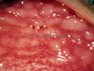
Clinical image of Follicular conjunctivitis - imageId=2897559. Click to open in gallery.  caption: 'Gray-white lymphoid follicles and surrounding dilated blood vessels on the tarsal conjunctiva, secondary to <i>Chlamydia</i> infection.'
Gray-white lymphoid follicles and surrounding dilated blood vessels on the tarsal conjunctiva, secondary to Chlamydia infection.
Copyright © 2023 VisualDx®. All rights reserved.