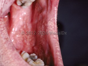
Fordyce spots are normal, superficial, ectopic sebaceous glands seen on mucosal surfaces that are not associated with hair follicles. They may affect as much as 80% of the population. Fordyce spots are often found on the oral mucosa, the vermilion of the lips, the inferior eyelids, and the genitals. The spots are usually clustered and asymptomatic.
The likelihood of developing Fordyce spots increases with age; however, these small papules can be seen in patients of any age. Their development can be associated with hormonal changes, and puberty can bring about their onset.
Fordyce spots - Oral Mucosal Lesion
See also in: Overview,AnogenitalAlerts and Notices
Important News & Links
Synopsis

Codes
ICD10CM:
L73.9 – Follicular disorder, unspecified
Q38.6 – Other congenital malformations of mouth
SNOMEDCT:
50584008 – Fordyce's disease
L73.9 – Follicular disorder, unspecified
Q38.6 – Other congenital malformations of mouth
SNOMEDCT:
50584008 – Fordyce's disease
Look For
Subscription Required
Diagnostic Pearls
Subscription Required
Differential Diagnosis & Pitfalls

To perform a comparison, select diagnoses from the classic differential
Subscription Required
Best Tests
Subscription Required
Management Pearls
Subscription Required
Therapy
Subscription Required
References
Subscription Required
Last Reviewed:03/29/2021
Last Updated:04/11/2021
Last Updated:04/11/2021
 Patient Information for Fordyce spots - Oral Mucosal Lesion
Premium Feature
VisualDx Patient Handouts
Available in the Elite package
- Improve treatment compliance
- Reduce after-hours questions
- Increase patient engagement and satisfaction
- Written in clear, easy-to-understand language. No confusing jargon.
- Available in English and Spanish
- Print out or email directly to your patient
Upgrade Today

Fordyce spots - Oral Mucosal Lesion
See also in: Overview,Anogenital