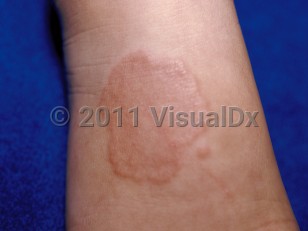
Granuloma annulare (GA) is a common inflammatory skin disorder of unknown etiology involving the dermis or subcutaneous tissues. The disease is seen in children and adults, and incidence peaks in the third or fourth decade of life. The condition is more common in girls than boys.
Several variants of GA exist, including localized (the most common), generalized, subcutaneous, perforating, macular, vesicular, palmoplantar, and blaschkolinear variants. Generalized forms of GA are more chronic or persistent than localized forms. Palmoplantar GA can be painful. The subcutaneous variant occurs almost exclusively in children and favors the lower extremities.
Human leukocyte antigen (HLA)-Bw35 has been associated with generalized GA and thyroid disease.
GA may have a prolonged course, but lesions tend to spontaneously resolve without scarring.
Granuloma annulare in Infant/Neonate
See also in: External and Internal EyeAlerts and Notices
Important News & Links
Synopsis

Codes
ICD10CM:
L92.0 – Granuloma annulare
SNOMEDCT:
65508009 – Granuloma annulare
L92.0 – Granuloma annulare
SNOMEDCT:
65508009 – Granuloma annulare
Look For
Subscription Required
Diagnostic Pearls
Subscription Required
Differential Diagnosis & Pitfalls

To perform a comparison, select diagnoses from the classic differential
Subscription Required
Best Tests
Subscription Required
Management Pearls
Subscription Required
Therapy
Subscription Required
Drug Reaction Data
Subscription Required
References
Subscription Required
Last Reviewed:10/12/2020
Last Updated:10/12/2020
Last Updated:10/12/2020
 Patient Information for Granuloma annulare in Infant/Neonate
Premium Feature
VisualDx Patient Handouts
Available in the Elite package
- Improve treatment compliance
- Reduce after-hours questions
- Increase patient engagement and satisfaction
- Written in clear, easy-to-understand language. No confusing jargon.
- Available in English and Spanish
- Print out or email directly to your patient
Upgrade Today

Granuloma annulare in Infant/Neonate
See also in: External and Internal Eye