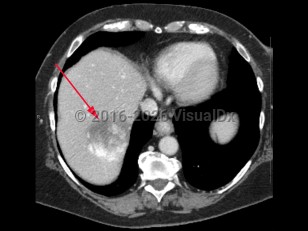
Hepatic hemangiomas, also known as cavernous hemangiomas, are common benign mesenchymal tumors of the liver. The majority of hepatic hemangiomas are solitary and can range in size from a few millimeters to over 20 cm (giant hemangiomas). Prevalence of hepatic hemangiomas range from 0.4%-20%, with diagnosis most frequently between the third and fifth decade of life.
Most hepatic hemangiomas are asymptomatic and are incidental findings on abdominal imaging. Larger lesions are more likely to cause symptoms, including abdominal pain (especially in the upper right quadrant), nausea, and anorexia. Women are 3 times as likely to develop hemangiomas and are more likely to experience symptoms. The etiology of hepatic hemangiomas is unclear, but hormonal influence over tumor enlargement may explain the higher incidence of symptomatic lesions in women. Lesions may grow during pregnancy and in the setting of oral contraceptive therapy. Surgical resection may be necessary for patients experiencing pain.
Hepatic hemangioma
Alerts and Notices
Important News & Links
Synopsis

Codes
ICD10CM:
D18.09 – Hemangioma of other sites
SNOMEDCT:
93469006 – Hemangioma of liver
D18.09 – Hemangioma of other sites
SNOMEDCT:
93469006 – Hemangioma of liver
Look For
Subscription Required
Diagnostic Pearls
Subscription Required
Differential Diagnosis & Pitfalls

To perform a comparison, select diagnoses from the classic differential
Subscription Required
Best Tests
Subscription Required
Management Pearls
Subscription Required
Therapy
Subscription Required
Drug Reaction Data
Subscription Required
References
Subscription Required
Last Reviewed:01/07/2018
Last Updated:01/07/2018
Last Updated:01/07/2018
Hepatic hemangioma