Immunoglobulin A vasculitis (IgAV), formerly called Henoch-Schönlein purpura, is a necrotizing small-vessel vasculitis of unknown etiology that is the most common form of vasculitis in children aged younger than 10 years. It is characterized by IgA-immune complex and C3 and fibrin deposition in small vessels: primarily capillaries, postcapillary venules, and occasionally arterioles in affected organs. The annual incidence in childhood is 3-26 per 100 000. It is seen more frequently in males, White individuals, and those of Asian descent, commonly during fall and winter seasons. Episodes can often be preceded by an upper respiratory infection.
IgAV is characterized clinically by palpable purpura, abdominal pain, arthritis, and hematuria. Children are more likely to have fevers and abdominal pain. There are prodromal symptoms of malaise, headache, and arthralgias. An individual episode may persist for 3-6 weeks, with recurrences in many patients.
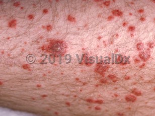
Almost all patients develop palpable purpura. Other skin involvement may include petechiae, bullae, edema, and necrosis. Infants' skin involvement is sometimes limited to the face or arms. Joint abnormalities are the second most common symptom and may accompany skin eruptions with severe pain and sometimes swelling, warmth, and tenderness. Ankles and knees are most often involved; symptoms are often transient and migratory.
Severe abdominal pain, vomiting, hematemesis, diarrhea, and hematochezia occur in about 50% of children. An involved inflamed and edematous bowel may be accompanied by appendicitis, ileus, and infarction. Intussusception and bowel perforation are more common in children than in adults.
Renal involvement occurs in 20%-50% of children and is usually self-limited, with only 1%-3% of patients progressing to end-stage renal disease. Risk factors for more severe renal disease include increased age at presentation, increased levels of serum IgA, and baseline nephritic or nephrotic disease. Notably, skin lesions above the waist may indicate higher risk in adults, but studies have not shown this in children.
Immunoglobulin A vasculitis in Infant/Neonate
Alerts and Notices
Important News & Links
Synopsis

Codes
ICD10CM:
D69.0 – Allergic purpura
SNOMEDCT:
191306005 – Henoch-Schönlein purpura
D69.0 – Allergic purpura
SNOMEDCT:
191306005 – Henoch-Schönlein purpura
Look For
Subscription Required
Diagnostic Pearls
Subscription Required
Differential Diagnosis & Pitfalls

To perform a comparison, select diagnoses from the classic differential
Subscription Required
Best Tests
Subscription Required
Management Pearls
Subscription Required
Therapy
Subscription Required
Drug Reaction Data
Subscription Required
References
Subscription Required
Last Updated:09/27/2022
Immunoglobulin A vasculitis in Infant/Neonate