Keratoacanthoma in All Ages
See also in: External and Internal Eye,Hair and ScalpAlerts and Notices
Important News & Links
Synopsis

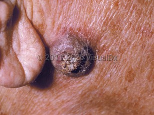
Keratoacanthoma is a rapidly growing, well-differentiated neoplasm of squamous epithelium. Many consider keratoacanthomas to be a low-grade variant of squamous cell carcinoma (SCC). In contrast to classic SCCs, keratoacanthomas typically appear and grow rapidly over a few weeks and spontaneously involute and resolve within 6 months, leaving an atrophic scar. They are most commonly seen in individuals aged 60 years and older with light skin colors and a history of prolonged sun exposure. Men are more commonly affected than women. Risk factors include UV radiation, human papillomavirus infection, immunosuppression, and certain medications. Patients on immunosuppressant medications tend to have more persistent and chronic keratoacanthomas.
Codes
ICD10CM:
L85.8 – Other specified epidermal thickening
SNOMEDCT:
254662007 – Keratoacanthoma
L85.8 – Other specified epidermal thickening
SNOMEDCT:
254662007 – Keratoacanthoma
References
Subscription Required
Last Updated:03/29/2026
 Patient Information for Keratoacanthoma in All Ages
Premium Feature
VisualDx Patient Handouts
Available in the Elite package
- Improve treatment compliance
- Reduce after-hours questions
- Increase patient engagement and satisfaction
- Written in clear, easy-to-understand language. No confusing jargon.
- Available in English and Spanish
- Print out or email directly to your patient
Upgrade Today

Keratoacanthoma in All Ages
See also in: External and Internal Eye,Hair and Scalp