A keratoacanthoma is a rapidly growing, well-differentiated neoplasm of squamous epithelium. In contrast to classic squamous cell carcinomas (SCC), keratoacanthomas typically appear and grow rapidly over a few weeks and spontaneously involute and resolve within 6 months, leaving an atrophic scar. The immune system is thought to play a role in the spontaneous regression of keratoacanthomas.
Keratoacanthomas are most commonly seen in individuals aged 60 years and older with lighter skin colors and a history of prolonged sun exposure. Men are more commonly affected than women.
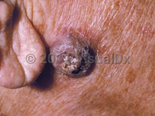
Keratoacanthomas typically present as solitary, crater-shaped nodules measuring a couple centimeters in diameter, often with a central keratin plug on sun-exposed skin.
A periocular keratoacanthoma occurs around the eye and grows rapidly over the course of a few weeks to months. Lesions that involve the eye may produce mechanical abnormalities, such as ectropion or ptosis, and, occasionally, may cause destructive changes. If left untreated, some periocular keratoacanthomas spontaneously involute within 6 months, but they may lead to scarring and destruction of ocular adnexa.
Risk factors for keratoacanthomas include ultraviolet radiation, human papillomavirus infection, immunosuppression, and certain medications. Patients on immunosuppressant medications tend to have more persistent and chronic keratoacanthomas. Patients taking medications such as BRAF inhibitors or hedgehog inhibitors have also been reported to develop keratoacanthomas. In addition, skin injury may be a predisposing factor, and there are reports of keratoacanthomas developing in sites of prior trauma, in surgical scars, after laser resurfacing, and following radiation therapy. In rare cases, keratoacanthomas may develop as part of a syndrome.
Many consider keratoacanthomas to be a low-grade variant of SCC. Most will cause only local destruction. Due to the very thin skin of the eyelid, periocular lesions are particularly susceptible to extension into underlying stroma and even orbicularis oculi muscle. More invasive variants, with metastasis to draining lymph nodes or the cavernous sinus, have been reported.
Keratoacanthoma - External and Internal Eye
See also in: Overview,Hair and ScalpAlerts and Notices
Important News & Links
Synopsis

Codes
ICD10CM:
L85.8 – Other specified epidermal thickening
SNOMEDCT:
254662007 – Keratoacanthoma
L85.8 – Other specified epidermal thickening
SNOMEDCT:
254662007 – Keratoacanthoma
Look For
Subscription Required
Diagnostic Pearls
Subscription Required
Differential Diagnosis & Pitfalls

To perform a comparison, select diagnoses from the classic differential
Subscription Required
Best Tests
Subscription Required
Management Pearls
Subscription Required
Therapy
Subscription Required
Drug Reaction Data
Subscription Required
References
Subscription Required
Last Reviewed:03/06/2024
Last Updated:03/18/2024
Last Updated:03/18/2024
 Patient Information for Keratoacanthoma - External and Internal Eye
Premium Feature
VisualDx Patient Handouts
Available in the Elite package
- Improve treatment compliance
- Reduce after-hours questions
- Increase patient engagement and satisfaction
- Written in clear, easy-to-understand language. No confusing jargon.
- Available in English and Spanish
- Print out or email directly to your patient
Upgrade Today

Keratoacanthoma - External and Internal Eye
See also in: Overview,Hair and Scalp