Lentigo maligna, historically referred to as a Hutchinson melanotic freckle, is the most common subtype of melanoma in situ, accounting for about 80% of cases. It primarily affects older adults, most often in the sixth and seventh decades of life, and arises on chronically ultraviolet (UV) radiation-exposed areas of the head and neck, particularly in individuals with Fitzpatrick skin phototype I to II.
Lentigo maligna is a slow-growing, noninvasive proliferation of atypical intraepidermal melanocytes. While some sources distinguish between lentigo maligna and melanoma in situ, lentigo maligna type (the latter of which is thought to be more malignant), the World Health Organization (WHO) recognizes lentigo maligna and melanoma in situ as the same entity.
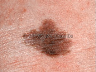
The natural history of lentigo maligna is that of gradual, asymmetric, radial growth. The majority of lesions are larger than 6 mm and present as flat macules or patches with ill-defined, irregular borders. Pigmentation is variegated, ranging from light brown to black and sometimes including areas of hypopigmentation suggestive of partial regression. Lentigo maligna has a particular predilection for the nose and cheeks.
The lesion typically remains in the radial growth phase for many years, with melanocytic proliferation confined to the basal layer and lateral spread. Approximately 5% progress to lentigo maligna melanoma, marked by vertical growth, with a mean time to progression of about 28 years. Clinical signs of progression include the development of a palpable nodule, increased pigmentation, and bleeding.
Lentigo maligna and lentigo maligna melanoma are associated with chronic sun damage and may occur in patients with a history of nonmelanoma skin cancers. Additional associations include immunosuppression, Werner syndrome, oculocutaneous albinism, and xeroderma pigmentosum.
Lentigo maligna
See also in: Hair and ScalpAlerts and Notices
Important News & Links
Synopsis

Codes
ICD10CM:
D03.9 – Melanoma in situ, unspecified
SNOMEDCT:
302836005 – Lentigo maligna
D03.9 – Melanoma in situ, unspecified
SNOMEDCT:
302836005 – Lentigo maligna
Look For
Subscription Required
Diagnostic Pearls
Subscription Required
Differential Diagnosis & Pitfalls

To perform a comparison, select diagnoses from the classic differential
Subscription Required
Best Tests
Subscription Required
Management Pearls
Subscription Required
Therapy
Subscription Required
References
Subscription Required
Last Reviewed:04/30/2026
Last Updated:05/03/2026
Last Updated:05/03/2026
 Patient Information for Lentigo maligna
Premium Feature
VisualDx Patient Handouts
Available in the Elite package
- Improve treatment compliance
- Reduce after-hours questions
- Increase patient engagement and satisfaction
- Written in clear, easy-to-understand language. No confusing jargon.
- Available in English and Spanish
- Print out or email directly to your patient
Upgrade Today

Lentigo maligna
See also in: Hair and Scalp