Lichen sclerosus (LS) is a chronic dermatosis that primarily involves the anogenital area. It is characterized by an initial inflammatory phase that is often followed by changes to genital architecture and skin atrophy if left untreated. There are 2 reported peaks of onset, the first in premenarchal girls and the second in postmenopausal women, but it is thought that onset during the reproductive years is likely underrecognized.
The pathogenesis of LS is unknown, but an autoimmune mechanism with genetic predisposition is hypothesized, with an increased prevalence of HLA-DQ7, DQ8, DQ9, and DR12. Some studies implicate Borrelia spp and Epstein-Barr virus (EBV) infection, and trauma and hormonal influences have been investigated. LS has also been reported in the setting of immune checkpoint inhibitor therapy.
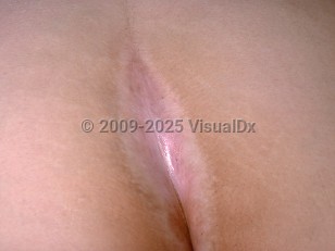
Over 85% of lesions are found on anogenital skin. Characteristic sites include the clitoris and clitoral hood, interlabial sulci, labia minora, and perineum. The medial labia majora and perianal area may also be involved. LS of the vaginal mucosa is exceedingly rare.
LS can progress to cause agglutination of labia minora to adjacent mucosa, and phimosis of the clitoral hood to the clitoris. Sclerosis of the vaginal introitus can also occur. Difficulty in voiding may be seen in the later stages.
The main symptom is pruritus, and at times, this can be incapacitating, interfering with daily activity and sleep. Alternatively, LS can be asymptomatic in a smaller percentage of patients. Secondary erosion or ulceration may lead to burning and pain, especially after micturition. Dyspareunia may occur secondary to active perineal disease or introital narrowing.
Complications include secondary lichen simplex chronicus and secondary infection. Superimposed allergic contact dermatitis may develop. Squamous cell carcinoma (SCC) can occur within LS genital lesions. This may be preceded by differentiated vulvar intraepithelial neoplasia (dVIN). There is a higher risk of SCC in untreated cases. Furthermore, several case series describe an association between vulvar melanoma and LS.
Around 10% percent of women with vulvar LS have extragenital LS.
Lichen sclerosus - Anogenital in
See also in: Overview,External and Internal EyeSynopsis

Codes
ICD10CM:
L90.0 – Lichen sclerosus et atrophicus
SNOMEDCT:
895454001 – Lichen sclerosus
L90.0 – Lichen sclerosus et atrophicus
SNOMEDCT:
895454001 – Lichen sclerosus
Look For
Subscription Required
Diagnostic Pearls
Subscription Required
Differential Diagnosis & Pitfalls

To perform a comparison, select diagnoses from the classic differential
Subscription Required
Best Tests
Subscription Required
Management Pearls
Subscription Required
Therapy
Subscription Required
Drug Reaction Data
Subscription Required
References
Subscription Required
Last Reviewed:07/08/2025
Last Updated:07/27/2025
Last Updated:07/27/2025
 Patient Information for Lichen sclerosus - Anogenital in
Premium Feature
VisualDx Patient Handouts
Available in the Elite package
- Improve treatment compliance
- Reduce after-hours questions
- Increase patient engagement and satisfaction
- Written in clear, easy-to-understand language. No confusing jargon.
- Available in English and Spanish
- Print out or email directly to your patient
Upgrade Today

Lichen sclerosus - Anogenital in
See also in: Overview,External and Internal Eye