This summary discusses adult patients. Linear IgA bullous dermatosis of childhood is addressed separately.
Linear IgA bullous dermatosis (LABD) is a rare immune-mediated disorder that occurs in both children and adults. The disease is defined by the presence of a homogenous deposition of IgA in a linear band at the epidermal-dermal junction. The 3 main types of LABD include the drug-induced, pediatric, and adult forms of the disease.
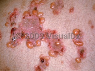
In the adult form, LABD most commonly presents in the fifth decade of life. Clinical presentation is highly variable, ranging from urticarial plaques, papules, and papulovesicles to large erosions, and the rash can be focal or generalized. Mucosal erosions (which can affect any mucosal surface) are common and may be severe and result in scarring. LABD may be associated with ulcerative colitis and, rarely, with hematologic and solid organ malignancies.
The drug-induced LABD variant usually occurs 1-4 weeks after starting the offending medication and resolves within several weeks of stopping the medication. It resembles classic LABD clinically. It can present with more widespread bullous involvement than non-drug-induced disease. Localized or widespread macular erythema may be seen in association. Localized, morbilliform, erythema multiforme-like with targetoid lesions, and Stevens-Johnson-like presentations have also been described. A number of drugs have been associated with LABD, most commonly vancomycin. Other causative agents include a variety of antibiotics (eg, trimethoprim-sulfamethoxazole, amoxicillin), NSAIDs, simvastatin, cyclosporine, furosemide, lithium, captopril, phenytoin, interleukin-2, interferon gamma, and amiodarone. IgA nephropathy can be rarely associated with the cutaneous eruption of drug-induced LABD.
Most patients with LABD (of any type) have IgA1 antibodies to a 97-kDa and a 120-kDa fragment of the extracellular portion of bullous pemphigoid antigen 2 (BP180 / type XVII collagen), typically the 15 collagenous domain and less frequently the NC16A epitope. Reactivity to collagen VII, laminin-332, and laminin gamma-1 have also been described. Collagen VII has been shown to be the antigen target in a small group of patients with vancomycin-induced LABD.
Linear IgA bullous dermatosis
Alerts and Notices
Important News & Links
Synopsis

Codes
ICD10CM:
L13.8 – Other specified bullous disorders
SNOMEDCT:
95330001 – Linear immunoglobulin A dermatosis
L13.8 – Other specified bullous disorders
SNOMEDCT:
95330001 – Linear immunoglobulin A dermatosis
Look For
Subscription Required
Diagnostic Pearls
Subscription Required
Differential Diagnosis & Pitfalls

To perform a comparison, select diagnoses from the classic differential
Subscription Required
Best Tests
Subscription Required
Management Pearls
Subscription Required
Therapy
Subscription Required
Drug Reaction Data
Subscription Required
References
Subscription Required
Last Reviewed:02/10/2020
Last Updated:03/08/2020
Last Updated:03/08/2020
Linear IgA bullous dermatosis