Livedoid vasculopathy
Alerts and Notices
Important News & Links
Synopsis

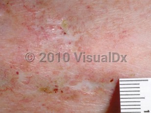
Livedoid vasculopathy (LV), also known as livedoid vasculitis, presents with retiform purpura or ulcers on the lower legs that may progress to atrophie blanche (AB). LV is caused by vaso-occlusion of the cutaneous microcirculation that manifests histopathologically as hyalinization of vessels leading to fibrin thrombi. LV is a rare condition, with an estimated incidence of 1:100 000 per year and an increased incidence during summer months and during pregnancy. It tends to affect young to middle-aged adults, with a median age of onset of 30-50 years, and has a female-to-male ratio of 2-3:1.
The pathogenesis of LV is not well understood, but it is likely related to increased coagulability and/or decreased fibrinolysis. The strongest risk factor is a hypercoagulable state with laboratory evaluation positive for a known cause of thrombophilia in 40%-50% of patients in retrospective cohort studies. The second most common risk factor is connective tissue disease. LV can also be associated with monocolonal gammopathy, with hepatitis B or C, or can be idiopathic.
LV follows a chronic time course, with lesions often developing for months before a definitive diagnosis is made. In a recent case series, the median interval between symptom onset and diagnosis was 10 months and between onset and treatment was 22.5 months.
The condition is rare in children.
Codes
ICD10CM:
L95.0 – Livedoid vasculitis
SNOMEDCT:
238762002 – Livedoid vasculitis
L95.0 – Livedoid vasculitis
SNOMEDCT:
238762002 – Livedoid vasculitis
Look For
Subscription Required
Diagnostic Pearls
Subscription Required
Differential Diagnosis & Pitfalls
- Chronic venous insufficiency with AB
- Chilblains – Usually a history of cold exposure.
- Erythema ab igne – Look for history of chronic heat source exposure.
- Lichen sclerosus – Look for shiny, atrophic plaques on the anogenital regions (often in a figure-of-eight pattern) and trunk.
- Localized scleroderma (morphea) – Look for ivory, indurated plaques on other areas of the body, often with a violaceous rim.
- Malignant atrophic papulosis – Look for smaller lesions on the trunk or proximal extremities, surrounded by a distinctive telangiectatic rim.
- Livedo reticularis, physiologic – Look for presence upon cold exposure and resolution with rewarming.
- Livedo reticularis, idiopathic – This is a diagnosis of exclusion. The condition is persistent, with no known triggers. It is more common in young women and exaggerated upon cold exposure.
- Livedo reticularis, primary – This is a diagnosis of exclusion. Lesions appear and disappear spontaneously without known triggers.
- Livedo reticularis, autoimmune connective tissue disorders – Look for evidence of systemic lupus erythematosus, antiphospholipid syndrome, vasculitis, dermatomyositis, etc.
- Livedo reticularis, hematologic disorders – Look for cryoglobulinemia, deep vein thrombosis, disseminated intravascular coagulation.
- Livedo reticularis, embolic or deposition – Look for dyslipidemia, septic emboli, calciphylaxis (end-stage renal failure).
- Livedo reticularis, drug-induced – Look for history of minocycline, amantadine, quinidine, etc.
- Ulcers, arterial – Look for history of arterial disease. Very painful punched-out ulcers occur on the feet, toes, or heels. Buerger disease with a long, extensive smoking history is very common.
- Ulcers, venous – Look for history of extensive varicose veins, chronic venous stasis, and ulcers on the inner portion of the lower leg.
- Ulcers, neuropathic – Look for history of diabetes or neuropathy.
- Ulcers, traumatic – Look for history of trauma.
- Ulcers, vasculitis – Leukocytoclastic vasculitis and polyarteritis nodosa may have ulcerations as part of their presentation.
- Calciphylaxis – Commonly on fat-bearing areas of the body but can present on the lower extremities. Characterized by exquisitely painful retiform purpura.
Best Tests
Subscription Required
Management Pearls
Subscription Required
Therapy
Subscription Required
References
Subscription Required
Last Reviewed:06/03/2020
Last Updated:06/04/2020