Contents

SynopsisCodesLook ForDiagnostic PearlsDifferential Diagnosis & PitfallsBest TestsManagement PearlsTherapyDrug Reaction DataReferences

Information for Patients

View all Images (35)

Other Resources UpToDate PubMed

Lobular capillary hemangioma in Child

See also in: External and Internal Eye,Hair and Scalp,Nail and Distal Digit,Oral Mucosal Lesion
Contributors: Jeffrey M. Cohen MD, Belinda Tan MD, PhD, Susan Burgin MD
Other Resources UpToDate PubMed

Synopsis

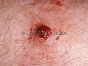
Lobular capillary hemangiomas, also known as pyogenic granulomas, are rapidly growing, usually single, benign vascular growths. The etiology is unknown. These lesions may arise within other vascular malformations, such as larger hemangiomas or superficial telangiectases. Trauma may also precipitate these lesions. When more than one pyogenic granuloma is present, they may be clustered together, or agminated.

They can bleed profusely after even minor trauma. While they can occur in patients aged younger than 6 months, they generally occur in older children. A 2004 study reported an average age of 5.9 years. Lobular capillary hemangiomas comprise 0.5% of all skin nodules in children.

Codes

ICD10CM:
L98.0 – Pyogenic granuloma

SNOMEDCT:
200722003 – Pyogenic granuloma

Look For

Subscription Required

Diagnostic Pearls

Subscription Required

Differential Diagnosis & Pitfalls

Best Tests

Subscription Required

Management Pearls

Subscription Required

Therapy

Subscription Required

Drug Reaction Data

Below is a list of drugs with literature evidence indicating an adverse association with this diagnosis. The list is continually updated through ongoing research and new medication approvals. Click on Citations to sort by number of citations or click on Medication to sort the medications alphabetically.

Subscription Required

References

Subscription Required

Last Reviewed:11/05/2017
Last Updated:05/07/2019
Copyright © 2023 VisualDx®. All rights reserved.
Patient Information for Lobular capillary hemangioma in Child
Print E-Mail Images (35)
Contributors: Medical staff writer

Overview

Lobular capillary hemangioma, also known as pyogenic granuloma, is a common non-cancerous (benign) growth that often appears as a rapidly growing, bleeding bump on the skin or inside the mouth. It is made up of blood vessels and may occur at a location that has had a minor injury.

Who’s At Risk

Lobular capillary hemangiomas occur in people of all races. Women are more frequently affected than men, though male and female children are equally affected.

A lobular capillary hemangioma is most often seen in:
  • Children and young adults
  • Pregnant women
  • Women taking oral contraceptives
  • People taking certain oral retinoids, including isotretinoin or acitretin (Soriatane)
  • People taking protease inhibitors such as indinavir (Crixivan)
  • People undergoing chemotherapy

Signs & Symptoms

The most common locations for a lobular capillary hemangioma include:
  • Hands, fingers, and forearms
  • Head, face, and neck
  • Lips, gums, and inner mouth (particularly in pregnant women)
  • Trunk
Usually, a lobular capillary hemangioma appears as a beefy, red bump that enlarges rapidly over a few weeks. On average, a lobular capillary hemangioma is 5-10 mm in diameter. It is likely to bleed easily, and, in some cases, can be tender to the touch. Rarely, multiple lesions of lobular capillary hemangiomas may develop at the same time (simultaneously) at the same location.

Self-Care Guidelines

Any rapidly growing skin growth should be examined by a physician in order to confirm the correct diagnosis. Because lobular capillary hemangiomas may bleed easily, cover the lesion with a bandage until you see your child's doctor.

When to Seek Medical Care

Make an appointment with your child's doctor or a dermatologist if any rapidly growing or bleeding growth develops on your child's skin or in the areas lining body cavities (mucous membranes) such as the mouth, nose, ears, eyes, or genitalia.

Treatments

If the diagnosis of lobular capillary hemangioma is suspected, the doctor may perform a skin biopsy. The procedure involves:
  1. Numbing the skin with an injectable anesthetic.
  2. Sampling a small piece of skin by using a flexible razor blade, a scalpel, or a tiny cookie cutter (called a "punch biopsy"). If a punch biopsy is taken, a suture or two may be placed and will need to be removed 6-14 days later.
  3. Having the skin sample examined under the microscope by a specially trained physician (dermatopathologist).
Lobular capillary hemangiomas associated with medications usually shrink (regress) when the medicine is stopped or the dosage is lowered. Depending on the size of the lobular capillary hemangioma, location, and symptoms, the doctor may decide that no treatment is necessary for people who can safely stop or lower the dose of the triggering medication.

Although lobular capillary hemangioma is a harmless condition, it is frequently removed due to its tendency to bleed, its tenderness to the touch, and its distressing appearance. However, lobular capillary hemangiomas may go away on their own without treatment.

If the lobular capillary hemangioma is obvious, the physician may choose to treat it immediately after obtaining a biopsy. Such treatments include:
  • Scraping and burning (curettage and cauterization). After numbing with local anesthetic, the area is scraped with a sharp instrument (a curette) and burned with an electric needle (cautery)
  • Silver nitrate solution
  • Topical imiquimod cream (Aldara)
  • Laser treatment
  • Freezing with liquid nitrogen (cryotherapy)
  • Surgical removal (excision)
Approximately 40% of lobular capillary hemangiomas can come back after treatment, especially those lesions located on the trunk of teens and young adults. A lobular capillary hemangioma that comes back (recurs) is best treated by surgical removal.

References


Bolognia, Jean L., ed. Dermatology, pp.1823-1824. New York: Mosby, 2003.

Freedberg, Irwin M., ed. Fitzpatrick's Dermatology in General Medicine. 6th ed, pp.1003, 1009. New York: McGraw-Hill, 2003.
Copyright © 2023 VisualDx®. All rights reserved.
Lobular capillary hemangioma in Child
See also in: External and Internal Eye,Hair and Scalp,Nail and Distal Digit,Oral Mucosal Lesion
A medical illustration showing key findings of Lobular capillary hemangioma : Developed acutely , Friable papule, Hemorrhagic skin lesion, Red color
Clinical image of Lobular capillary hemangioma - imageId=163970. Click to open in gallery.  caption: 'A close-up of a well-demarcated bleeding papule.'
A close-up of a well-demarcated bleeding papule.
Copyright © 2023 VisualDx®. All rights reserved.