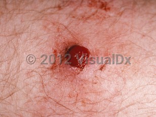
Lobular capillary hemangioma, also known as pyogenic granuloma, is a tumor-like, benign reactive, vascular proliferation, essentially an "-oma" of granulation tissue, usually caused by trauma or local irritation. It is not a true neoplastic process. It does not usually suppurate so the term "pyogenic" is a misnomer.
It tends to occur in children and young adults, and 75% occur on the gingiva. However, the labial mucosa, buccal mucosa, and tongue may also be affected. Over time, lobular capillary hemangiomas may sclerose and become more fibrous.
A particular subset occurs in pregnant patients, and this is called "granuloma gravidarum." These occur in the first trimester of pregnancy and grow steady under hormonal influences throughout pregnancy. Postpartum, the lesions tend to become smaller or involute completely. However, many will remain as a smaller, scarred, fibrotic nodule. Patients are aware of an enlarging mass that may bleed easily.
Lobular capillary hemangioma - Oral Mucosal Lesion
See also in: Overview,External and Internal Eye,Hair and Scalp,Nail and Distal DigitAlerts and Notices
Important News & Links
Synopsis

Codes
ICD10CM:
L98.0 – Pyogenic granuloma
SNOMEDCT:
200722003 – Pyogenic granuloma
L98.0 – Pyogenic granuloma
SNOMEDCT:
200722003 – Pyogenic granuloma
Look For
Subscription Required
Diagnostic Pearls
Subscription Required
Differential Diagnosis & Pitfalls

To perform a comparison, select diagnoses from the classic differential
Subscription Required
Best Tests
Subscription Required
Management Pearls
Subscription Required
Therapy
Subscription Required
Drug Reaction Data
Subscription Required
References
Subscription Required
Last Updated:05/08/2023
 Patient Information for Lobular capillary hemangioma - Oral Mucosal Lesion
Premium Feature
VisualDx Patient Handouts
Available in the Elite package
- Improve treatment compliance
- Reduce after-hours questions
- Increase patient engagement and satisfaction
- Written in clear, easy-to-understand language. No confusing jargon.
- Available in English and Spanish
- Print out or email directly to your patient
Upgrade Today

Lobular capillary hemangioma - Oral Mucosal Lesion
See also in: Overview,External and Internal Eye,Hair and Scalp,Nail and Distal Digit