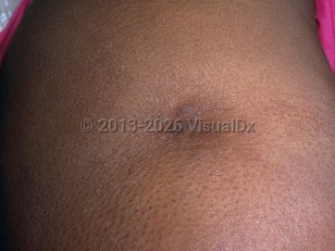
Lupus panniculitis is a form of chronic cutaneous lupus erythematosus characterized by firm subcutaneous nodules and plaques. Although the lesions of lupus panniculitis can involve the face, upper arms, trunk, buttocks, and legs, there is a predilection for facial involvement in the pediatric population. The affected areas tend to be asymptomatic and may or may not have associated overlying cutaneous changes. The lesions resolve over time, resulting in subcutaneous atrophy that can be disfiguring.
This entity is rare in the pediatric population, and its exact prevalence is unknown. The mean age of onset is 8 years among the cases reported. Most patients do not have concurrent systemic lupus erythematosus (SLE), and extensive laboratory evaluation is not indicated in the absence of systemic symptoms.
Lupus panniculitis in Child
Alerts and Notices
Important News & Links
Synopsis

Codes
ICD10CM:
L93.2 – Other local lupus erythematosus
SNOMEDCT:
239888002 – Lupus panniculitis
L93.2 – Other local lupus erythematosus
SNOMEDCT:
239888002 – Lupus panniculitis
Look For
Subscription Required
Diagnostic Pearls
Subscription Required
Differential Diagnosis & Pitfalls

To perform a comparison, select diagnoses from the classic differential
Subscription Required
Best Tests
Subscription Required
Management Pearls
Subscription Required
Therapy
Subscription Required
References
Subscription Required
Last Reviewed:05/26/2025
Last Updated:05/27/2025
Last Updated:05/27/2025
Lupus panniculitis in Child