Contents

SynopsisCodesLook ForDiagnostic PearlsDifferential Diagnosis & PitfallsBest TestsManagement PearlsTherapyDrug Reaction DataReferences

Information for Patients

View all Images (3)

Melanoma - Oral Mucosal Lesion
See also in: Overview,Anogenital,Hair and Scalp
Other Resources UpToDate PubMed

Melanoma - Oral Mucosal Lesion

See also in: Overview,Anogenital,Hair and Scalp
Contributors: Reba Suri MD, Carl Allen DDS, MSD, Belinda Tan MD, PhD, Sook-Bin Woo MS, DMD, MMSc, Susan Burgin MD
Other Resources UpToDate PubMed

Synopsis

Although melanoma is classically thought of as a cutaneous malignancy, it can also arise from the mucosal epithelium of the respiratory, alimentary, and genitourinary systems. While cutaneous melanoma is well studied, the literature on mucosal melanoma is limited. This synopsis focuses specifically on melanoma of the oral cavity.

Melanoma is a rare malignancy of the oral cavity. It accounts for around 0.5% of all melanomas. Black individuals and individuals of Japanese descent are affected more often. There is a 2:1 male predilection, and most occur in the sixth decade of life or later.

Oral melanoma can present as a painless, bleeding mass or ulcer of the hard palate or maxillary gingiva, but about 60% of cases are asymptomatic and identified incidentally during a dental examination. It tends to be diagnosed when advanced, and only approximately one-third of patients give a history of a pre-existing pigmented lesion, either nevus or mucosal melanosis. The risk factors for oral melanoma are not well studied. Smoking, irritation from dentures, and alcohol use have been proposed but not confirmed as risk factors. Only around 10%-38% of patients survive 5 years or more.

Related topic: nail melanoma

Codes

ICD10CM:
C43.9 – Malignant melanoma of skin, unspecified

SNOMEDCT:
372244006 – Malignant melanoma

Look For

Subscription Required

Diagnostic Pearls

Subscription Required

Differential Diagnosis & Pitfalls

  • Benign melanocytic nevus
  • Amalgam tattoo (exogenous pigmentation) – This is the most common intraoral pigmented lesion. Usually the amalgam tattoo is not elevated and particles of dental amalgam (silver filling material) can often be detected on periapical radiographs of the lesional site.
  • Melanotic macule / post-traumatic melanosis – These focal areas of melanin deposition could appear clinically identical to a junctional melanocytic nevus. Biopsy would be necessary to distinguish between the two.
  • Melanoacanthoma – This uncommon condition occurs most frequently in young adult Black women, typically on the buccal mucosa. The lesion is generally much larger (1 cm or greater) than a melanocytic nevus, and it often follows a characteristic pattern of spontaneous involution over a period of days to weeks.
  • Blue nevus is usually less than 1 cm in size and evenly pigmented.
  • Physiologic pigmentation may appear ominous to a patient if they had not noticed the extensive pigmentation in the past. The pigment is even throughout and symmetric in distribution.
  • Drug-induced pigmentation may have a sudden onset and rapid progression, but the pigment is even and there is no nodularity. This may be particularly concerning in patients infected with HIV.
  • Lobular capillary hemangioma (pyogenic granuloma) may grow rapidly and bleed but tend to not arise from a macule pigmented area.
  • Kaposi sarcoma may appear similar; a biopsy differentiates between the two.
  • Oral squamous cell carcinoma
  • Malignant salivary gland and soft tissue tumors

Best Tests

Subscription Required

Management Pearls

Subscription Required

Therapy

Subscription Required

Drug Reaction Data

Below is a list of drugs with literature evidence indicating an adverse association with this diagnosis. The list is continually updated through ongoing research and new medication approvals. Click on Citations to sort by number of citations or click on Medication to sort the medications alphabetically.

Subscription Required

References

Subscription Required

Last Reviewed:01/22/2022
Last Updated:01/27/2022
Copyright © 2023 VisualDx®. All rights reserved.
Patient Information for Melanoma - Oral Mucosal Lesion
Print E-Mail Images (3)
Contributors: Medical staff writer
Premium Feature
VisualDx Patient Handouts
Available in the Elite package
  • Improve treatment compliance
  • Reduce after-hours questions
  • Increase patient engagement and satisfaction
  • Written in clear, easy-to-understand language. No confusing jargon.
  • Available in English and Spanish
  • Print out or email directly to your patient
Copyright © 2023 VisualDx®. All rights reserved.
Melanoma - Oral Mucosal Lesion
See also in: Overview,Anogenital,Hair and Scalp
A medical illustration showing key findings of Melanoma : Black color, Pigmented papule
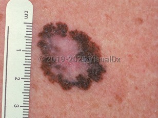
Clinical image of Melanoma - imageId=732728. Click to open in gallery.  caption: 'A close-up of a large variegated plaque with pink, dark brown, and rust colors. Note also irregular borders and asymmetry.'
A close-up of a large variegated plaque with pink, dark brown, and rust colors. Note also irregular borders and asymmetry.
Copyright © 2023 VisualDx®. All rights reserved.