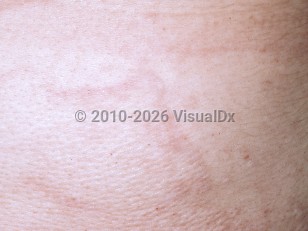
Mid-dermal elastolysis (MDE) is a rare acquired disorder characterized by the idiopathic loss of mid-dermal elastic fibers. MDE typically presents with asymptomatic, well-demarcated thin wrinkled plaques on the upper arms and trunk (type I). Preceding erythema is reported by a minority of patients and, even more rarely, urticarial plaques may be seen. Variant presentations of MDE include soft plaques with perifollicular protrusions with a peau d'orange appearance (type II) and reticular erythema (type III).
Most reported patients are women of Northern European descent between the ages of 30 and 50 years. Men with MDE have a slightly older age of onset. Systemic involvement is not a feature.
The pathogenesis of this condition has not been fully elucidated; however, purported pathomechanisms include ultraviolet light-induced elastic fiber degeneration, defects in the synthesis of elastic fibers, and autoimmune destruction thereof.
Mid-dermal elastolysis
Alerts and Notices
Important News & Links
Synopsis

Codes
ICD10CM:
L94.8 – Other specified localized connective tissue disorders
SNOMEDCT:
238821003 – Idiopathic mid-dermal elastolysis
L94.8 – Other specified localized connective tissue disorders
SNOMEDCT:
238821003 – Idiopathic mid-dermal elastolysis
Look For
Subscription Required
Diagnostic Pearls
Subscription Required
Differential Diagnosis & Pitfalls

To perform a comparison, select diagnoses from the classic differential
Subscription Required
Best Tests
Subscription Required
Management Pearls
Subscription Required
Therapy
Subscription Required
References
Subscription Required
Last Updated:06/06/2016
Mid-dermal elastolysis