Pediatric
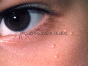
Milia (singular, milium) are minute epidermoid cysts (also known as infundibular cysts) that present as small white or yellow papules, usually on the face. They are typically smaller than 3 mm in diameter. They occur when sloughed off skin becomes trapped in the dermis. There is no predilection for either sex or for any race or ethnicity.
Primary milia affect 40%-50% of newborns but may be found in patients of all ages. Secondary milia often occur after cosmetic procedures (dermabrasion, chemical peels, ablative laser therapy) or trauma, or in conjunction with a number of blistering disorders. Milia may occur in tattoos. Patients with skin phototypes IV through VI tend to be more likely to develop milia as sequelae of chemical peels. Blistering disorders that may heal with milia and scarring include epidermolysis bullosa acquisita; porphyrias, including porphyria cutanea tarda; bullous pemphigoid; herpes zoster; contact dermatitis (allergic, irritant); bullous lupus erythematosus; and dermatitis herpetiformis. Milia have also been known to occur in areas of topical steroid-induced atrophy. Persistent or widespread milia are associated with a number of syndromes (see Differential Diagnosis & Pitfalls).
Codes
ICD10CM: L72.8 – Other follicular cysts of the skin and subcutaneous tissue
SNOMEDCT: 254679001 – Milia
Look For
Subscription Required
Diagnostic Pearls
Subscription Required
Differential Diagnosis & Pitfalls
Milia are sometimes confused with closed comedones of acne
Below is a list of drugs with literature evidence indicating an adverse association with this diagnosis. The list is continually updated through ongoing research and new medication approvals. Click on Citations to sort by number of citations or click on Medication to sort the medications alphabetically.
Milia are commonly found on the skin of people of all ages. They are formed when keratin (a substance produced by the skin) becomes entrapped beneath the outer layer of the skin, forming a tiny cyst. An individual milium (the singular of milia) is formed at the base of a hair follicle or sweat gland.
Milia can be categorized as either primary or secondary. Primary milia are formed directly from entrapped keratin and are usually found on the faces of infants and adults. Secondary milia are also tiny cysts and look similar, but these develop after something clogs the ducts leading to the skin surface, such as after an injury, burn, or blistering of the skin.
Who’s At Risk
Milia can occur in people of all ages, of any ethnicity, and of either sex. Milia are so common in newborn babies (occurring in up to 50% of them) that they are considered normal.
Secondary milia may appear in affected skin of people with the following:
Blistering skin conditions, such as bullous pemphigoid, epidermolysis bullosa, and porphyria cutanea tarda
Burns
Blistering injury to skin, such as poison ivy
Following skin resurfacing procedures such as dermabrasion or laser resurfacing
Following long-term use of steroid creams
Chronic sun damage
Signs & Symptoms
Milia appear as 1–2 mm white-to-yellow, dome-shaped bumps that are not painful or itchy.
The most common locations for primary milia include:
Around the eyes, cheeks, nose, and forehead in adults and infants
On the gums and palate inside the mouth of infants; these milia are called Epstein's pearls, and they occur in up to 85% of infants
The most common locations for secondary milia include:
Anywhere on the body where another skin condition exists, particularly on the backs of the hands
On the faces of people who have had a lot of damage from sun exposure
Self-Care Guidelines
Primary milia found in infants tend to heal on their own within several weeks, though the primary milia found in adults tend to be long-lasting.
Although milia are found in the outer layers of skin, they are difficult to remove without the proper tools. Do not try to remove them at home, as you may leave a scar.
When to Seek Medical Care
See a dermatologist or your general physician for evaluation if you notice any new bumps on the skin.
Treatments
If the diagnosis from the doctor is primary milia in an infant, no treatment is necessary, as the bumps will go away on their own within a few weeks.
Primary or secondary milia in an adult may disappear on their own, but your doctor may treat you with one of the following:
Piercing each milium with a sterile lancet or scalpel followed by removal of the cyst material with a tool called a comedone extractor
Topical retinoid cream such as tretinoin, tazarotene, or adapalene
A series of acid peels or microdermabrasion procedures at the dermatologist's office
References
Bolognia, Jean L., ed. Dermatology, pp.1722-1723. New York: Mosby, 2003.
Freedberg, Irwin M., ed. Fitzpatrick's Dermatology in General Medicine. 6th ed. pp.601, 604, 780. New York: McGraw-Hill, 2003.