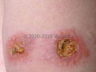
Necrobiotic xanthogranuloma (NXG) is a rare histiocytic disorder that presents as firm yellow papules and plaques of the skin, most often in the periorbital area. It is strongly associated with an immunoglobulin G (IgG) monoclonal gammopathy (80%-90% of cases), particularly IgG kappa (60%). NXG may affect extracutaneous tissues including the eye, spleen, heart, lung, kidney, intestine, ovary, larynx, pharynx, skeletal muscle, and central nervous system. Ophthalmologic complications occur in about 50%-80% of patients, and may include orbital masses, conjunctival involvement, keratitis, scleritis, and uveitis.
In a 2020 multicenter study and systematic review of 235 cases of NXG, the mean age at presentation was 61.8 years with a 3:2 female to male predominance.
NXG often follows a protracted and indolent course, but patients with the condition also have an increased risk of plasma cell dyscrasias (multiple myeloma and monoclonal gammopathy of undetermined significance [MGUS]) and other lymphoproliferative disorders. When these coexisting conditions exist, the disease may follow a fatal course.
The etiology of NXG is unknown. It is speculated that the paraprotein, present in the majority of cases, binds to lipoprotein receptors of monocytes, inducing formation of a xanthogranuloma, or may bind to serum lipoproteins.
Necrobiotic xanthogranuloma
Alerts and Notices
Important News & Links
Synopsis

Codes
ICD10CM:
D76.3 – Other histiocytosis syndromes
SNOMEDCT:
404164003 – Necrobiotic xanthogranuloma
D76.3 – Other histiocytosis syndromes
SNOMEDCT:
404164003 – Necrobiotic xanthogranuloma
Look For
Subscription Required
Diagnostic Pearls
Subscription Required
Differential Diagnosis & Pitfalls

To perform a comparison, select diagnoses from the classic differential
Subscription Required
Best Tests
Subscription Required
Management Pearls
Subscription Required
Therapy
Subscription Required
References
Subscription Required
Last Reviewed:05/31/2020
Last Updated:05/31/2020
Last Updated:05/31/2020
Necrobiotic xanthogranuloma